 English
 Français
 Español
 Deutsch
 Italiano
 العربية


Tel :+86-755-86961139
Email :sales@vivo-light.com
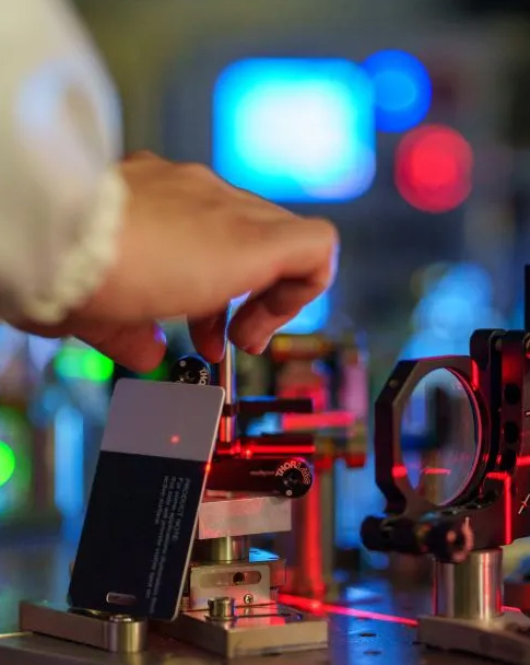

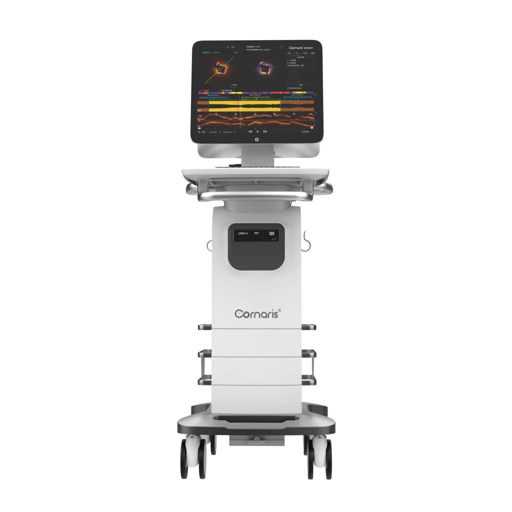
You see a transformation in cardiology with intravascular OCT imaging equipment. Advanced systems like the Cornaris P80 give you sharp optical coherence tomography images, helping you see vessel details and guide interventions with confidence. Recent studies show that OCT-guided PCI changes treatment strategies for many patients and improves patient outcomes.
The number of OCT-guided PCIs increased by 548.4% from 2011 to 2019.
The ILUMIEN I study found that optical coherence tomography influenced PCI strategies in 57% of cases.
|
Imaging Modality |
Benefits |
|---|---|
|
Optical Coherence Tomography (OCT) |
Enhanced plaque characterization |
|
Intravascular Ultrasound (IVUS) |
Detailed structural insights |
|
Intravascular Photoacoustic Imaging (IVPA) |
Compositional and biomechanical insights |
You experience a new era in cardiovascular medicine with intravascular oct imaging equipment. This technology uses optical coherence tomography to deliver high-resolution images inside coronary arteries. The system sends near-infrared light through a catheter, which penetrates vessel walls and reflects back detailed information. You see individual tissue layers and microfeatures with clarity. Coherence gating and optical interferometry allow you to detect single-scattered light, producing images with a resolution of 10 to 15 micrometers. Rapid imaging speeds, reaching up to 200 frames per second, help you visualize coronary arteries in real time during percutaneous coronary intervention.
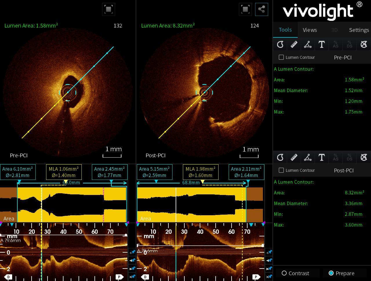
The Cornaris P80 stands out in cardiology. You benefit from its multimodality approach, which combines Index of Plaque Attenuation, Virtual Flow Ratio, and Intelligent Calcium Assessment. These features let you assess plaque stability, measure functional flow, and identify calcium severity—all in one pullback. You save time and avoid extra procedures. The system offers multiple imaging modes, including Full View Mode for long pullbacks and High-definition Mode for ultra HD scans. You adapt to different clinical applications with ease.
|
Mechanism |
Contribution to Imaging Capabilities |
|---|---|
|
High Spatial Resolution |
Enables optimal imaging of individual tissue layers and microfeatures of therapeutic devices. |
|
Near-Infrared Light |
Provides deeper penetration (1 to 3 mm) into highly scattering media, enhancing image quality. |
|
Coherence Gating |
Allows detection of single-scattered light, generating high-resolution images (10 to 15 µm). |
|
Rapid Imaging Speeds |
Facilitates high frame rates (150 fps or more), improving clinical imaging of coronary arteries. |
|
Optical Interferometry |
Offers superior sensitivity compared to traditional methods, detecting backscattered signals with high dynamic range. |
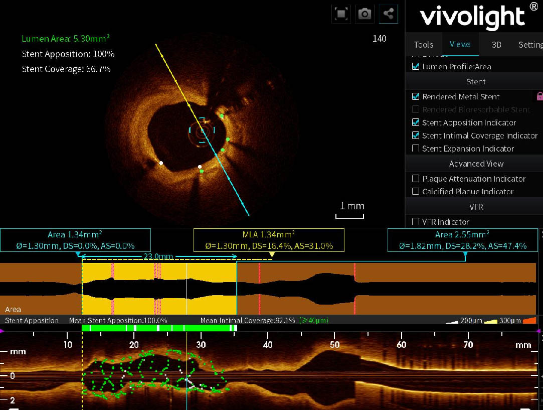
You use intravascular imaging to vividly delineate vessel wall pathology, including plaque morphology and composition. The technology visualizes stent apposition and tissue coverage at an individual strut level. You see calcified plaques without artifacts, measure calcium distribution, and assess plaque thickness. These capabilities improve diagnosis and treatment planning in clinical practice.
|
Feature/Capability |
Cornaris P80 (Multimodality) |
Single-Modality OCT Systems |
|---|---|---|
|
Integration of Technologies |
Combines multiple advanced technologies in one platform |
Limited to one imaging modality |
|
Comprehensive Assessments |
Allows for detailed assessments without extra procedures |
Requires additional procedures |
|
Unique Diagnostic Features |
Includes Index of Plaque Attenuation, Virtual Flow Ratio, Intelligent Calcium Assessment |
Lacks advanced functional assessments |
|
Preoperative Functional Assessments |
Provides critical insights for treatment strategies |
Limited preoperative insights |
|
Efficiency |
Single pullback for multimodal results |
Multiple pulls needed for different assessments |
You see direct heart health benefits when you use optical coherence tomography during percutaneous coronary intervention. Intravascular imaging guidance leads to better clinical outcomes for patients. You reduce major adverse cardiac events, cardiac death, and stent thrombosis. The technology helps you identify vulnerable plaque features, such as thin caps and macrophage infiltration. You optimize stent placement and minimize complications.
You improve patient outcomes by reducing all-cause mortality, myocardial infarctions, and target lesion revascularization. The following table summarizes reductions in major cardiac events observed in clinical trials:
|
Study/Trial |
Outcome |
Reduction (%) |
|---|---|---|
|
LANCET Network Meta-Analysis |
Target Lesion Failure |
24% |
|
LANCET Network Meta-Analysis |
Cardiac Death |
39% |
|
LANCET Network Meta-Analysis |
Stent Thrombosis |
53% |
|
RENOVATE-COMPLEX-PCI trial |
Major Adverse Cardiac Events |
7.7% vs. 12.3% (HR 0.64; P=0.008) |
|
Network Meta-Analysis |
All-cause Death |
45% |
|
Network Meta-Analysis |
All-cause Myocardial Infarction |
25% |
|
OCTOBER trial |
Major Adverse Cardiac Events |
28% |
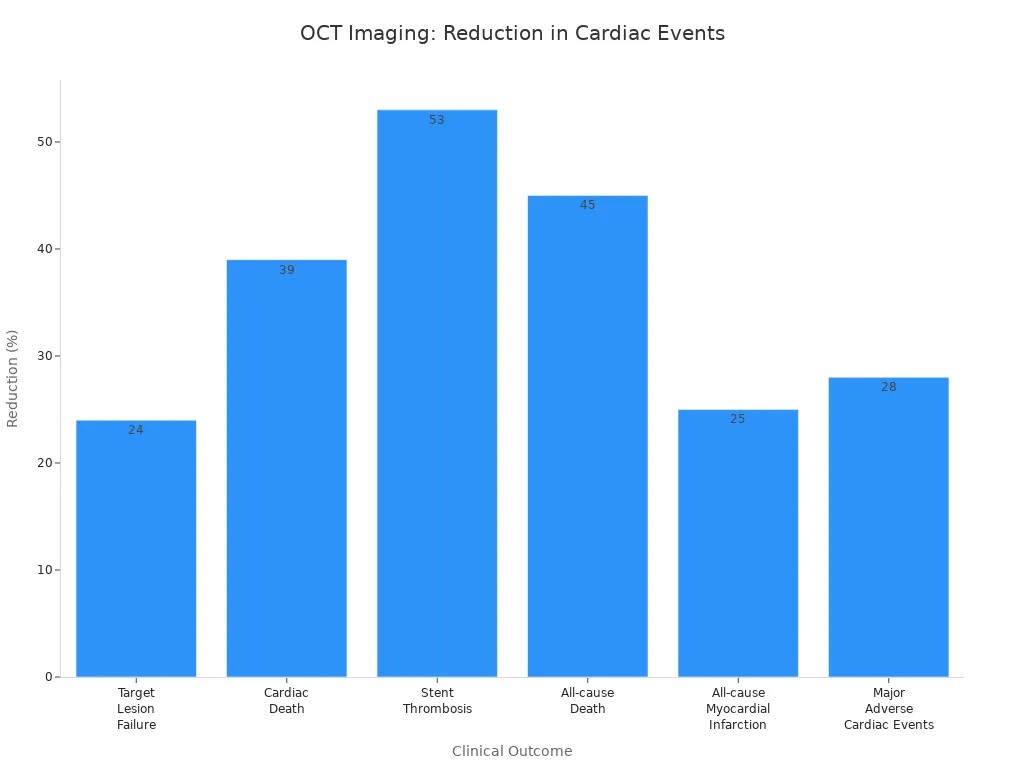
You observe these improvements in clinical outcomes across multiple trials:
The RENOVATE-COMPLEX-PCI trial showed a significant reduction in major adverse cardiac events with imaging guidance compared to angiography-guided PCI.
OCT-guided PCI demonstrated a 28% reduction in major adverse cardiac event rates in complex bifurcation lesions as per the OCTOBER trial.
You use intravascular oct imaging equipment to guide percutaneous coronary intervention and improve long-term outcomes for patients. Intravascular imaging guidance led to a 25% reduction in all-cause mortality. Cardiac death decreased by 45%. Myocardial infarctions dropped by 17%. Stent thrombosis was reduced by 48%. Target vessel myocardial infarction fell by 18%. Target lesion revascularization decreased by 28%.
You rely on optical coherence tomography for vulnerable plaque detection and plaque characterization. The technology provides high-resolution images that allow for detailed characterization of plaques. You identify vulnerable features and enhance procedural guidance during interventions. You see improvements in the management of coronary artery disease and clinical applications in cardiovascular medicine.
Recent developments in catheter design have improved maneuverability in complex cases. Faster acquisition rates reduce motion artifacts, leading to clearer images. The flexibility of intravascular imaging in clinical settings optimizes the management of coronary artery disease and supports better diagnosis and treatment strategies.
You gain a powerful advantage in cardiology when you use intravascular OCT imaging equipment for diagnosis. High-resolution imaging lets you see the smallest details inside coronary arteries. You can visualize tissue layers, detect minor defects, and assess neointimal hyperplasia thickness with precision. This level of detail helps you identify issues that other imaging methods might miss.
You can quantify neointimal hyperplasia and assess its characteristics.
You detect small tissue prolapse and edge dissections with greater accuracy than with IVUS.
The depth of penetration, less than 500 micrometers, makes optical coherence tomography ideal for evaluating specific patient populations.
|
Evidence Description |
Key Findings |
|---|---|
|
High-resolution imaging from OCT |
Produces accurate automated quantification of vessel lumen dimensions and higher sensitivity in identifying plaque characteristics and morphology. |
|
Comparison with IVUS |
Demonstrates superior ability to visualize and characterize plaques, assess vessel size, and detect small morphological changes post-stenting. |
You use these capabilities to improve diagnosis and guide clinical decisions. Automated calculations and multiple imaging modes, such as Full View and High-definition, help you adapt to different clinical scenarios. Preprocedural imaging with optical coherence tomography often changes your original percutaneous coronary intervention strategy. In fact, you may alter your plan in over 80% of cases after reviewing OCT images.
You achieve greater precision in treatment when you rely on intravascular imaging. Optical coherence tomography provides accurate quantification of coronary calcification, which is essential for stent expansion and reducing complications. You can assess the location, extent, and thickness of calcium, information not available through traditional angiography.
You visualize arterial layers and detect even minor intimal thickening.
You distinguish between different atherosclerotic plaque components, such as fibrous, lipidous, and calcified tissues.
You identify thin cap fibroatheroma, which is critical for assessing vulnerable plaque detection.
|
Evidence Type |
Findings |
|---|---|
|
Improved Stent Expansion |
7% increase in Minimum Stent Area (MSA) with OCT guidance compared to angiography alone. |
|
Reduced Risk of Future Blockages |
64% reduction in stent thrombosis rates with OCT guidance. |
|
Additional Safety Benefits |
39% reduction in cardiac death and 24% reduction in target vessel myocardial infarction. |
You use these insights to select the best stenting strategies and adjunctive therapies. The technology helps you prevent geographic miss during stent placement, improving the effectiveness of your interventions. Real-time 3D visualization enhances your understanding of complex coronary lesions and supports personalized treatment strategies.
You see a clear reduction in procedural complications when you use intravascular OCT imaging equipment. Clinical trials show that optical coherence tomography guidance lowers the rates of stent thrombosis, major adverse cardiac events, and other complications during percutaneous coronary intervention.
|
Study |
Outcome Description |
Result |
|---|---|---|
|
ILUMIEN IV |
Reduction in stent thrombosis rates |
0.5% vs 1.4%, p=0.02 |
|
OCTOBER |
Reduced major adverse cardiac events (MACE) |
28% reduction compared to angiography |
|
OCTIVUS |
Fewer major procedural complications in OCT group |
2.2% vs 3.7% |
|
LANCET Meta-Analysis |
Significant reduction in all-cause death and MI |
45% reduction in death, 25% in MI |
You help patients achieve better clinical outcomes and long-term outcomes by minimizing risks during procedures. The integration of advanced imaging features, such as automatic calculations and 3D visualization, supports your efforts to deliver safer and more effective care. You use intravascular imaging to guide percutaneous coronary intervention, improve patient outcomes, and advance clinical applications in cardiovascular medicine.
You see the impact of intravascular oct imaging equipment in real-world cardiology through many success stories. In catheterization labs, you can combine advanced coronary reconstruction (ACR) with optical coherence tomography to improve major adverse cardiac event rates for patients with complex lesions. You notice that using optical coherence tomography often leads to changes in preprocedural strategies, which enhances treatment outcomes over a one-year follow-up.
You find that the HF-OCT catheter can reach extremely narrow lesions that standard catheters cannot access.
You benefit from greater flexibility when navigating tortuous arterial pathways, which is essential for complex coronary interventions.
You scan a wider range of vessel diameters, making treatment more effective for patients with challenging anatomy.
These advances in intravascular imaging help you deliver better care and improve long-term outcomes for patients with coronary artery disease.
You compare optical coherence tomography to other intravascular imaging methods and see clear differences in accuracy, speed, and safety. When you measure lumen area, you find that FD-OCT provides precise results with less variability than IVUS.
|
Measurement Type |
FD-OCT (mm²) |
IVUS (mm²) |
Variability (mm²) |
|---|---|---|---|
|
Lumen Area |
7.45 ± 0.17 |
8.03 ± 0.58 |
0.32 vs. 0.16 |
You also see that optical coherence tomography offers higher diagnostic accuracy for plaque characterization and vulnerable plaque detection.
|
Characterization Type |
OCT Sensitivity |
OCT Specificity |
IB-IVUS Sensitivity |
IB-IVUS Specificity |
C-IVUS Sensitivity |
C-IVUS Specificity |
|---|---|---|---|---|---|---|
|
Calcification |
100% |
100% |
100% |
99% |
100% |
100% |
|
Fibrosis |
98% |
94% |
94% |
84% |
93% |
93% |
|
Lipid Pool |
95% |
98% |
84% |
97% |
67% |
67% |
You experience faster imaging speeds with intravascular optical coherence tomography. Pullbacks reach 20-25 mm/s, while traditional angiography is much slower. You also see improved clinical outcomes in percutaneous coronary intervention, with lower risks of cardiac death and myocardial infarction. Intravascular imaging identifies critical lesion features that traditional methods may miss, supporting better treatment decisions and clinical applications in cardiovascular medicine.
You play a vital role in protecting patients during percutaneous coronary intervention. Intravascular oct imaging equipment gives you a clear view inside coronary arteries, which helps you avoid complications and improve safety. When you use optical coherence tomography, you follow a standardized workflow that reduces radiation exposure for both you and your patients. You do not see an increase in contrast utilization, which is important for patients with kidney concerns.
You minimize the need for universal vessel preparation in select lesions, which lowers the risk of unnecessary vessel trauma.
You increase the use of specialty devices, which helps you optimize stent implantation and reduce the chance of needing extra vessel treatment.
You enhance procedural efficiency, which means shorter procedure times and less stress for patients.
Clinical trials show that optical coherence tomography guidance leads to lower rates of major adverse cardiac events. Patients experience fewer complications, such as stent thrombosis, which boosts their confidence in the safety of the procedure.
|
Evidence Type |
Findings |
Patient Outcomes |
|---|---|---|
|
OCT-guided PCI |
Lower rates of major adverse cardiac events at 2 years (3.1% vs 4.9%) |
Increased patient satisfaction due to reduced complications |
|
Stent thrombosis |
Significantly lower in OCT-guided PCI |
Enhances patient confidence in procedure safety |
You also see that the use of intravascular imaging in complex cases does not increase in-hospital mortality. Standardized techniques help you handle challenging coronary lesions, which leads to better patient experiences and satisfaction.
You notice that optical coherence tomography transforms your workflow in the cardiac catheterization lab. Intravascular imaging provides automated, accurate measurements for stent selection, placement, and deployment. You save time because you do not need to repeat measurements or rely on guesswork.
|
Workflow Aspect |
Impact of Intravascular Imaging |
|---|---|
|
Measurement Accuracy |
Automated, precise measurements for stent selection |
|
Procedure Speed |
Faster workflows and reduced procedure times |
|
Adaptability |
Efficient handling of diverse clinical scenarios |
Many operators report that their workflows speed up when they use intravascular imaging technology. You adapt quickly to different clinical applications, whether you face simple or complex coronary artery disease. The integration of optical coherence tomography into your clinical practice supports better clinical outcomes and enhances your ability to deliver high-quality cardiovascular medicine. You rely on intravascular imaging for vulnerable plaque detection, plaque characterization, and improved patient care.
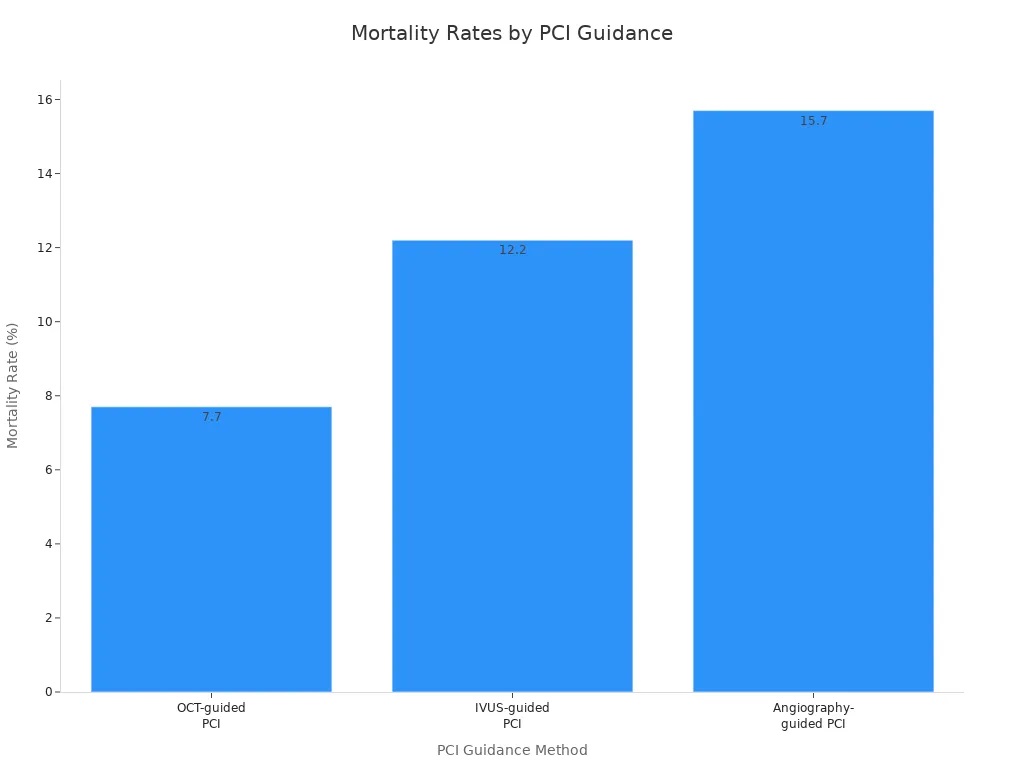
You see a clear connection between intravascular imaging and better heart health for patients in cardiology. Optical coherence tomography gives you higher procedural success and lower mortality rates compared to other imaging methods.
|
Group Type |
Mortality Rate (%) |
Procedural Success Rate |
MACE Rate |
|---|---|---|---|
|
OCT-guided PCI |
7.7 |
Higher |
Lower |
|
IVUS-guided PCI |
12.2 |
Moderate |
Moderate |
|
Angiography-guided PCI |
15.7 |
Lower |
Higher |
You benefit from advanced intravascular imaging features that improve clinical outcomes for coronary patients. Leading societies recommend optical coherence tomography for guiding coronary interventions and detecting stent issues. Intravascular imaging will continue to shape clinical practice as new technologies and applications emerge. You can help patients by adopting innovative intravascular imaging in cardiology.
Leave A Message
Scan to WhatsApp :


 IPv6 network supported