English
 Français
 Español
 Deutsch
 Italiano
 العربية


You can see how optical coherence tomography is now very important in heart care. Optical coherence tomography helps you see the heart’s arteries very clearly. Fast changes in optical coherence tomography keep making care better. These changes help you make good choices for your patients. Learning about new trends in oct technology helps get better results. Using multimodal oct imaging system changes how you check plaque stability. It also helps you do PCI better and follow new rules.

|
Evidence Type |
Description |
|---|---|
|
Performance |
The hybrid IVUS-OCT system works better than just IVUS or OCT alone for checking plaque and results after stent placement. |
|
Clinical Outcomes |
Studies show that using IVUS or OCT to guide PCI gives better results for patients. |
|
Guidelines Support |
The 2024 ESC Guidelines say IVI-guided PCI helps patients right away and in the future. |
Key Takeaways
-
Optical coherence tomography (OCT) gives clear pictures. These pictures help doctors see plaque and artery details. This helps doctors plan treatments better.
-
Multimodal OCT technology uses different imaging tools together. This helps doctors find heart disease early. It also helps patients get better results.
-
Ultra-fast OCT imaging makes the procedure quicker. This keeps patients safer. It lowers the chance of problems during heart procedures.
-
3D visualization in OCT helps doctors see complex artery shapes. This helps doctors place stents more accurately. It also leads to better treatment results.
-
Training and teamwork among healthcare workers are very important. This helps them use OCT technology well. It also helps improve patient care.
OCT Imaging System Advances
Multimodal OCT Technology
Today, you can use new optical coherence tomography systems. These systems put many imaging tools together in one place. Multimodal OCT technology lets you see more than just the outside of blood vessels. You can use optical coherence tomography, fundus photography, and fluorescein angiography at the same time. This helps you look deeper into the heart’s arteries and find problems early. Swept-source OCT and spectral-domain OCT give you clear pictures and more details about tissue. Multimodal OCT technology also uses AI to help you spot disease faster and make better choices.
|
Imaging Modality |
Advantages of Multimodal OCT |
Limitations of Traditional OCT |
|---|---|---|
|
Optical Coherence Tomography (OCT) |
High-resolution, cross-sectional images; integrates multiple imaging modalities |
Limited to single imaging type; lacks depth information |
|
Fundus Photography |
Widely used for 2D surface view |
No depth information; less detailed |
|
Fluorescein Angiography |
Effective for vascular abnormalities |
Invasive; risks allergic reactions |
|
Ultrasound B-scans |
Useful for posterior segment imaging |
Low resolution; not suitable for detailed analysis |
Now, you can use the oct imaging system to get different kinds of data in one scan. This makes your job quicker and more exact. Swept-source OCT and spectral-domain OCT help you see the heart’s arteries in new ways. You can use these tools to find issues before they get worse.
-
You can combine OCT, fundus photography, and fluorescein angiography.
-
This helps you find heart disease early and predict problems.
-
AI makes diagnosis better and faster.
High-Resolution Imaging
High-resolution imaging is very important in optical coherence tomography today. You can now see tiny details inside arteries that you could not see before. High-resolution images help you find thin-cap fibroatheromas and other signs of plaque that may cause trouble. Swept-source OCT and spectral-domain OCT both give you sharp images that show the layers of the artery wall. You can use these images to plan treatments and check if a stent is in the right spot.
|
Advancement Type |
Description |
|---|---|
|
Resolution |
OCT provides one to two orders of magnitude greater resolution than IVUS due to the smaller wavelength of light. |
|
Catheter Design |
The catheter for OCT can be much smaller than that used for IVUS, enhancing its usability. |
|
Non-Mechanical Scanning Systems |
Innovations in OCT technology are leading to the development of non-mechanical scanning systems, improving efficiency. |
With high-resolution imaging, you can see plaque features and measure how much calcium is in the artery. This helps you pick the best treatment for your patient. Clinical trials like ULTIMATE and ILUMIEN III show that high-resolution optical coherence tomography gives better results for patients who need PCI.
-
High-resolution OCT imaging shows plaque details and helps find weak spots.
-
OCT improves diagnosis by showing special plaque parts, which helps with risk checks and planning.
-
Studies like ULTIMATE and ILUMIEN III show that OCT-guided PCI works better than angiography-guided PCI.
Ultra-Fast OCT Imaging
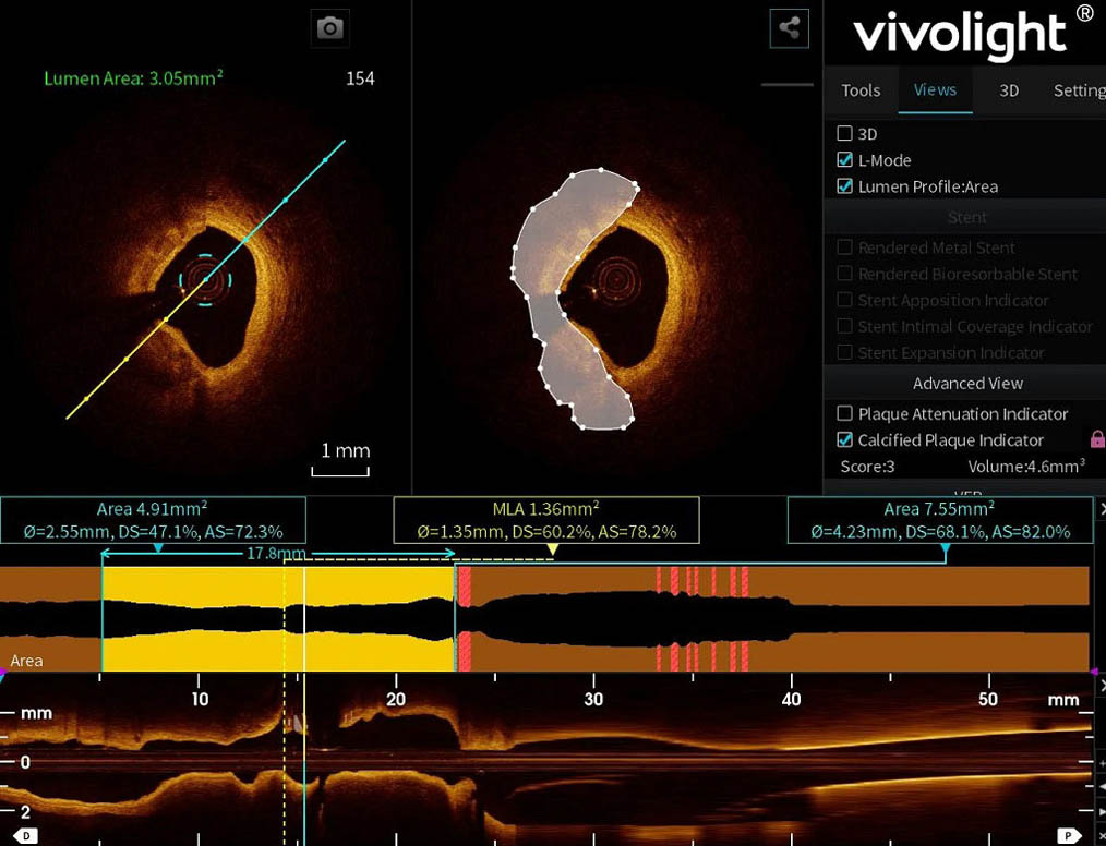
Ultra-fast optical coherence tomography changes how you work in the cath lab. You can now scan long parts of the artery in just a few seconds. Swept-source OCT and spectral-domain OCT both give fast imaging speeds. The Vivolight P80/P80-E system, for example, can scan 80 mm in only 2 seconds. This speed means you spend less time with the catheter in the artery, which lowers the risk of spasms and makes the procedure safer.
-
AI helps doctors read images faster and finish tasks sooner.
-
Both new and experienced doctors can work faster with AI.
-
OCT and AI give doctors quick answers and smart predictions.
-
These tools help treat heart disease sooner and more exactly.
-
The ultrafast imaging system works at 72 frames per second, so pictures are taken quickly and the procedure is shorter.
-
This speed keeps patients safer by lowering the chance of spasms.
-
Doctors can find dangerous plaques faster and plan better treatments.
You can use ultra-fast OCT imaging to get answers quickly and make choices right away. This helps you treat patients faster and feel more sure about your decisions.
3D Visualization
Three-dimensional visualization is a big improvement in optical coherence tomography. You can now see inside the artery from every side. 3D OCT helps you understand tricky places like where two arteries meet. Swept-source OCT and spectral-domain OCT both let you use 3D imaging. You can use 3D views to guide wires through stents and check if the stent fits well against the artery wall.
|
Benefit |
Description |
|---|---|
|
Enhanced Visualization |
3D OCT provides a clearer view of complex bifurcation anatomy compared to 2D imaging, aiding in the assessment of anatomical changes post-intervention. |
|
Clinical Application |
Quantitative measurements of side branch ostial area can be performed using 3D reconstructed OCT, facilitating the assessment of hemodynamic obstruction. |
|
Improved Guidewire Navigation |
3D reconstruction assists in guiding the re-crossing of guide wires through stent struts, optimizing the positioning and understanding of interactions between stents and vessel walls. |
Optical Coherence Tomography in Cardiology
Intravascular OCT Imaging
Doctors use optical coherence tomography to look inside blood vessels. This helps during heart procedures. Intravascular OCT imaging gives very clear pictures. You can see the layers of the artery wall. These images help find problems like plaque or stent issues. OCT shows much clearer pictures than intravascular ultrasound. You can see tiny details that ultrasound may not show.
-
In the United States, doctors used OCT in 0.3% of PCI procedures in 2014.
-
In Europe, OCT was used in 1.3% of PCI cases from 2005 to 2015.
-
In Japan, doctors used OCT in 11.6% of PCI procedures.
|
Feature |
Intravascular OCT |
Intravascular Ultrasound (IVUS) |
|---|---|---|
|
Resolution |
10–20 μm (high) |
100–150 μm (lower) |
|
Penetration Depth |
1–2 mm (limited) |
4–8 mm (greater) |
|
Plaque Characterization |
Detailed visualization of plaque types |
Useful for overall plaque morphology |
|
Detection of Complications |
Superior for edge dissections, stent malapposition |
Limited detection capabilities |
OCT imaging systems give high-resolution pictures. These help doctors make better choices during procedures.
Functional Assessment with OCT
Optical coherence tomography does more than show pictures. Doctors use OCT to measure how well blood moves through arteries. This helps decide if a blockage needs treatment. OCT imaging gives clear pictures and important blood flow data.
|
Clinical Outcome |
Description |
|---|---|
|
Improved prognosis |
Imaging-guided PCI shows better results than angiography-guided approaches, especially in tough cases. |
|
Functional assessment |
OCT-OFR gives both imaging and functional data, helping with planning and making procedures better. |
|
Tailored optimization |
After PCI, OCT-OFR can check results and let doctors make changes if needed. |
|
Evaluation of non-culprit lesions |
OCT-OFR helps guide full revascularization in cases with many vessel problems. |
|
In-stent restenosis evaluation |
OCT-OFR may help check ISR, but more studies are needed. |
Doctors use OCT to check if a stent works well. It also helps plan what to do next for the patient.
Plaque and Calcium Evaluation
Optical coherence tomography helps doctors find and study plaque and calcium in arteries. OCT gives very clear pictures of both calcified and non-calcified plaques. OCT is better than CT scans or ultrasound for finding small calcium spots. You can see the shape and size of plaque. This helps pick the best treatment.
-
Optical coherence tomography works well for finding and studying coronary plaque and calcium.
-
Doctors get detailed pictures of plaque parts, even tiny calcium spots.
-
OCT is better at showing plaque shapes, which helps with heart procedures.
Expanding OCT Clinical Applications
Optical coherence tomography has changed many heart procedures. Now, doctors use it for more things than before. The oct imaging system gives very clear pictures. It also helps guide and improve treatments. Let’s see how you can use optical coherence tomography for PCI, stent checks, and bifurcation lesions.
PCI Guidance
Doctors use optical coherence tomography to help with PCI. The clear images show the artery’s shape and help plan each step. You can see plaque, vessel size, and where to put stents. This makes the procedure safer and better.
New oct imaging lets you use different tools together. Some systems combine OCT with near-infrared spectroscopy. This helps you see plaque types without extra dye. Some systems use near-infrared fluorescence to show inflammation right away. You can spot dangerous plaques and pick the best treatment.
There are new features like IPA, OCT-FFR, and ICA. These give you more information from one scan. IPA checks if plaque is stable. OCT-FFR measures blood flow and shows if a blockage is bad. ICA uses AI to measure calcium and guide your treatment.
When you use optical coherence tomography for PCI, you lower the risk of problems. Studies show you get a bigger stent area and fewer issues like stent malapposition or edge dissection. You also see fewer problems in the hospital and better survival later.
You can see these benefits with new oct technology:
-
Multimodal systems help check plaque and guide PCI.
-
OCT-NIRS and near-infrared fluorescence show inflammation and risk.
-
Using OCT data with computer models gives better results.
Stent Optimization
You want every stent to work well and last long. Optical coherence tomography gives clear images of the stent inside the artery. You can check for gaps, tissue bulges, or poor expansion. This helps you fix problems right away.
The oct imaging system now has automatic tools for stent checks. You can use IPA to see if plaque is stable. OCT-FFR checks blood flow after putting in a stent. ICA measures calcium and helps treat tough spots. These tools save time and help you feel sure about your work.
|
Evidence Type |
Findings |
Impact |
|---|---|---|
|
Study by Ahmed et al. |
OCT-guidance made stent areas bigger (mean difference 0.35 mm²) |
Better stent results |
|
Cardiovascular mortality |
RR 0.56, 95% CI 0.32–0.99 |
Fewer deaths from heart problems |
|
Stent malapposition and major edge-dissection |
Happened less often |
Better outcomes for patients |
Optical coherence tomography helps you get better stent results. You lower the risk of stent clots and other problems. You also follow new rules that support OCT for finding artery disease.
Bifurcation Lesion Assessment
Treating bifurcation lesions is hard. You need to see both the main vessel and side branch. Optical coherence tomography gives clear, 3D images. You can see the vessel, wires, and stents in detail.
With oct imaging, you can:
-
Check the side branch opening and measure its size.
-
Pick the right stent size and where to put it.
-
Check wire positions and find broken struts.
-
Find clots and tears better than with IVUS.
You should check wire placement and stent position during the procedure. Clear out red blood cells for a better view. These steps help you get the best results.
The P80/P80-E system helps with bifurcation lesions. It gives automatic measurements and checks special stents. You get a full view and can spot risks early.
Now, doctors have more ways to use optical coherence tomography than ever. The oct imaging system gives clear images, many tools, and smart features. You can guide PCI, check stents, and treat bifurcation lesions more safely and successfully.
Research and Adoption Drivers
Clinical Evidence for OCT
Clinical evidence helps doctors use optical coherence tomography in heart care. Many studies show that OCT is a good tool for guiding heart procedures. Trials like ILUMIEN IV and OCTOBER compare OCT-guided ways with older methods. These studies show how OCT imaging makes patients safer and gives better results. Using the OCT imaging system helps doctors check arteries more accurately. It also helps pick the best treatment for each patient. With OCT technology, doctors can find artery problems early and help patients sooner.
Collaboration in Cardiology
Teamwork is important in heart care. Hospitals, device companies, and government groups work together. This teamwork helps everyone learn new ways to use optical coherence tomography. Sharing ideas and tools makes OCT technology get better faster. Working together gives doctors better tools and new OCT imaging systems for patients.
-
Big studies like ILUMIEN IV and OCTOBER test how well OCT-guided procedures work.
-
New AI models help doctors read OCT images more clearly.
-
Hospitals, companies, and government groups help OCT become more common.
-
Sharing tools and ideas helps make new OCT systems for doctors.
Training and Guidelines
Doctors need training to use optical coherence tomography well. Training programs teach doctors how to read OCT images and use them in care. Heart societies give rules for when to use OCT imaging in different cases. These rules help doctors use OCT technology safely and correctly. Following these rules helps doctors make better choices and helps patients get better.
Challenges and Future of OCT Imaging
Cost and Accessibility
Optical coherence tomography systems can cost a lot of money. Many hospitals have trouble buying these advanced tools. The price is usually between $40,000 and $150,000. Only big hospitals can pay for them. Small clinics and hospitals in poorer countries cannot afford these prices. This makes it hard for them to use optical coherence tomography for early heart checks.
|
Cost Range |
Implication for Adoption |
|---|---|
|
$40,000 - $150,000 |
High costs mean only big hospitals can buy them. |
|
$40,000 - $60,000 |
Too expensive for many screenings; prices must drop. |
|
Developing World |
Prices are too high for early heart checks. |
Some countries have more imaging centers and trained workers. Rich countries have better access to these systems. Poor countries have problems with cost, not enough equipment, and few trained people. New portable devices and better technology may help more places use optical coherence tomography soon.
Workflow Integration
Using oct in your clinic can take extra time. For example, oct-guided PCI may add about 18 minutes. You need special training to use these systems well. Some doctors worry about safety, like rare problems or using more contrast. As you learn and technology gets better, using oct will get easier.
-
You may need to teach your team how to read oct images.
-
Some hospitals must update their tools and rules.
-
In the long run, oct can save time by giving better information.
Regulatory Considerations
You must follow strict rules when you use optical coherence tomography. Each place has its own rules.
|
Region |
Regulatory Body |
Key Requirements |
|---|---|---|
|
EU |
Medical Device Regulation (MDR) 2017/745 |
CE marking, checking quality, watching safety after sales |
|
Japan |
Pharmaceuticals and Medical Devices Agency (PMDA) |
Review before sale, quality checks, safety after sale |
|
China |
National Medical Products Administration (NMPA) |
Device type, clinical checks, local tests |
|
Canada |
Health Canada |
Device license, quality checks, safety after sale |
You need to make sure your system is safe and passes tests. In the United States, you may need to talk to the FDA, check risk level, and send in forms. These steps help keep patients safe and make sure your oct system works right.
Future Innovations
New features will come soon for optical coherence tomography. Fourier-domain oct will make it easier to use and help doctors do more. You will get faster and better results with computer help and 3D color pictures. Artificial intelligence will help find disease, sort images, and watch changes over time. Deep learning models will make images clearer and help doctors make better choices.
You can see that optical coherence tomography is changing heart care. OCT gives doctors new tools to help make better choices. New advances in OCT give very clear pictures and quick answers. These things help patients get better and help doctors pick the right stent. When OCT systems use more than one tool, doctors get a full view. This helps them plan care that fits each patient. Experts think optical coherence tomography will keep getting more important. OCT will help doctors find and stop heart problems before they start.
-
Intracoronary OCT gives the same results when checking lesions.
-
OCT-guided PCI lowers the chance of problems and death.
-
Using more than one tool with OCT helps doctors make better choices.
As optical coherence tomography gets better, doctors will see more ways to help patients and find new ways to check for heart problems.

 IPv6 network supported