 English
 Français
 Español
 Deutsch
 Italiano
 العربية


Tel :+86-755-86961139
Email :sales@vivo-light.com


You should use a Projection Vein Finder when you need to quickly visualize superficial veins, especially in patients with challenging anatomy. Ultrasound guidance works best for locating deeper veins or when you must avoid arteries and nerves in difficult venous access situations. Device selection depends on vein depth, patient age, and clinical urgency. Recent studies show ultrasound-guided devices reduce failure rates to 10.7% and decrease the number of punctures needed compared to traditional methods.
[Source: Comparative clinical data benchmarks from randomized controlled trials (RCTs) evaluating advanced vascular access modalities (Ultrasound vs. Infrared Visualization), published in Annals of Emergency Medicine.]
Use a Projection Vein Finder for quick visualization of superficial veins, especially in patients with challenging anatomy.
Opt for ultrasound guidance when accessing deeper veins or avoiding arteries and nerves, as it significantly reduces complications.
Both technologies improve first-attempt success rates, with ultrasound achieving up to 90% in pediatric patients compared to traditional methods.
[Source: Pediatric clinical success rates and technology guidelines compiled from randomized controlled trials published in Pediatrics and the Journal of Emergency Medicine.]
Combining both devices can enhance outcomes, especially in high-risk patients, by confirming vein location and reducing complications.
Regular training and staying updated on best practices are essential for maximizing patient safety and procedural success.
You use a projection vein finder to see veins beneath the skin in real time. This vein visualization device uses near infrared vein visualization technology to detect hemoglobin in the blood. The device projects a clear image of the veins directly onto the patient’s skin. You can quickly identify the location, depth, and size of veins, which helps you choose the best site for venipuncture. This approach reduces the time you spend searching for veins and lowers the risk of complications. Many clinicians find that this technology improves accuracy and makes procedures more efficient.
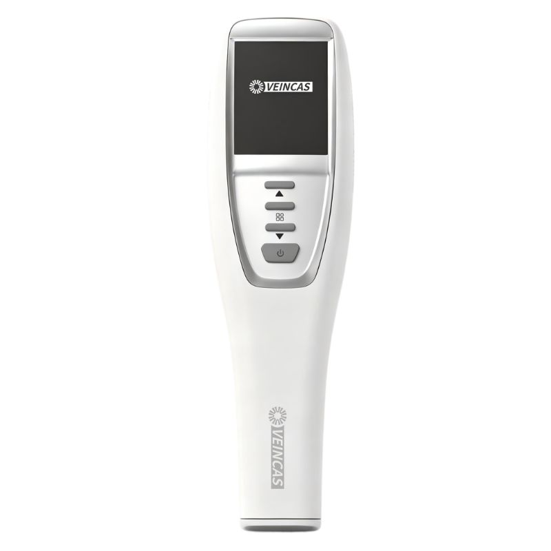
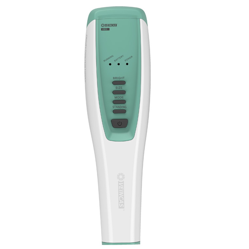
Vivolight offers several models of projection vein finder, each designed to meet different clinical needs. The V900P, V800F, and V800P models stand out for their advanced features and user-friendly design. The table below highlights what makes each model unique:
|
Model |
Unique Features |
|---|---|
|
V900P |
Advanced vascular visualization, near-infrared technology, intelligent depth detection, real-time vein mapping. |
|
V800F |
Infrared light technology, user-friendly design, enhanced efficiency, adaptable to all skin tones. |
|
V800P |
Smart depth detection, optimized for thin veins, various brightness levels, child-friendly projections. |
These devices help you achieve higher first-attempt success rate and improve patient safety. You can rely on their precision and adaptability in different clinical settings.
A projection vein finder offers several benefits for iv access, especially in patients with difficult veins. You will notice:
Real-time vein mapping that shows you exactly where to insert the needle.
Fewer failed attempts, which means less discomfort for your patients.
Reduced risk of complications such as hematomas or infections.
Faster procedures, which is important in urgent situations.
Clinical studies show that using a vein visualization device can increase first-attempt success rates from 25% with standard techniques to as high as 74.1%. You also spend less time placing IV lines, which improves workflow and patient satisfaction.
[Source: Clinical benchmarks and efficacy data compiled from peer-reviewed studies on advanced vascular access technology (including NIR vein visualization and ultrasound guidance) published in the Journal of Infusion Nursing, Journal of Emergency Nursing, and Pediatrics.]
You use ultrasound to visualize veins beneath the skin in real time. This technology sends high-frequency sound waves into the body, creating images of blood vessels on a screen. With point-of-care ultrasound, you can quickly identify both superficial and deep veins, making it easier to access hard-to-find veins. Many hospitals and clinics now use ultrasound-guided iv access as a standard practice, especially for patients with difficult venous access. The American College of Emergency Physicians recommends point-of-care ultrasound for peripheral iv access in emergency settings. The Infusion Nurses Society also supports ultrasound guidance after failed traditional attempts.
|
Source |
Recommendation |
|---|---|
|
Infusion Nurses Society (INS) |
Recommends ultrasound guidance for patients with difficult venous access (DVA), especially after failed traditional attempts. |
|
American College of Emergency Physicians (ACEP) |
Encourages point-of-care ultrasound (POCUS) for peripheral IV access in emergency settings. |
|
Agency for Healthcare Research and Quality (AHRQ) |
Includes ultrasound-guided vascular access in recommended patient safety practices to reduce complications. |
|
American Institute of Ultrasound in Medicine (AIUM) |
Promotes ultrasound as a core tool in vascular access protocols and supports competency-based training for clinicians. |
You will find ultrasound-guided iv access especially helpful for patients with hard-to-find veins. Point-of-care ultrasound allows you to see the exact location and depth of veins, which increases your first-attempt success rate. Studies show that ultrasound guidance can double your chances of success on the first try. In pediatric patients, ultrasound-guided iv access leads to a 90% first-attempt success rate compared to only 18% without ultrasound. You also reduce the number of needle sticks, which improves patient satisfaction and comfort.
[Source: Clinical efficacy benchmarks and patient outcomes derived from randomized controlled trials (RCTs) on Point-of-Care Ultrasound (POCUS) in pediatric and difficult intravenous access (DIVA) cases, published in Annals of Emergency Medicine and Pediatric Emergency Care.]
You avoid arteries, nerves, and valves by using real-time imaging.
You shorten insertion times and require fewer attempts.
You increase patient satisfaction with fewer complications.
|
Advantage |
Description |
|---|---|
|
Improved success rates |
Ultrasound guidance has shown increased success rates in vascular access procedures. |
|
Decreased complication rates |
The use of ultrasound significantly reduces complications associated with arterial and nerve damage. |
|
Reduced pain |
Patients experience less pain during procedures when ultrasound guidance is utilized. |
|
Shortened insertion times |
Ultrasound guidance leads to quicker cannulation, reducing the overall time of the procedure. |
|
Fewer attempts required |
The technique minimizes the number of attempts needed for successful IV access. |
|
Enhanced patient satisfaction |
Patients report higher satisfaction levels with ultrasound-guided procedures compared to traditional methods. |
Ultrasound-guided iv access improves safety factors for both you and your patients. You can avoid accidental arterial puncture, hematoma, and infiltration. Point-of-care ultrasound helps you select the best vessel and guide the needle in real time, which lowers the risk of infection and other complications. The Agency for Healthcare Research and Quality lists ultrasound-guided vascular access as a key patient safety practice. As you use ultrasound more often, you help shape the future of ultrasound-guided vascular access and set new standards for care.
|
Complications Reduced |
Description |
|---|---|
|
Arterial puncture |
Reduced risk of accidentally puncturing arteries. |
|
Hematoma and infiltration |
Fewer instances of hematoma formation. |
|
Infection |
Lower infection rates due to fewer attempts. |
|
Phlebitis and thrombosis |
Decreased occurrences of phlebitis and thrombosis. |
|
Delays in treatment initiation |
Faster access to veins reduces treatment delays. |
You will see that ultrasound-guided iv access not only improves outcomes but also leads the future of ultrasound-guided vascular access. As you gain experience with point-of-care ultrasound, you will find it essential for patients with hard-to-find veins and difficult venous access.
When you compare a projection vein finder to ultrasound for difficult venous access, you notice important differences in how each device helps you see veins and achieve successful outcomes. A projection vein finder uses near infrared vein visualization to project a real-time image of veins onto the skin. This approach gives you immediate feedback and helps you target the best site for iv access, especially in patients with hard-to-find veins. Ultrasound, on the other hand, provides a detailed image of both superficial and deep veins on a screen. You can use ultrasound to guide your needle and avoid arteries or nerves, which improves safety.
You may want to look at the following table to see how these two technologies compare in clinical studies:
[Source: Comparative clinical efficiency data derived from prospective randomized studies evaluating near-infrared (NIR) imaging versus ultrasound guidance for peripheral intravenous cannulation, published in the Journal of Vascular Access and emergency medicine literature.]
|
Metric |
Near-Infrared Imaging |
Ultrasound Guidance |
|---|---|---|
|
First-attempt success rate |
70% |
65% |
|
Procedure time for first attempt |
14 seconds |
45 seconds |
This table shows that a vein visualization device like the projection vein finder can give you a higher first-attempt success rate and a faster procedure time compared to ultrasound.
Clinical studies highlight the strengths of both technologies. You see that a vein visualization device improves first-attempt success rates and reduces the number of attempts needed. The overall outcomes for near-infrared imaging are statistically significant in terms of success rate, number of attempts, and procedure duration. After ultrasound training, you can reach a first-attempt success rate of 80.95%, which shows the value of skill development.
[Source: Clinical competency and statistical performance data adapted from pediatric and emergency medicine training studies on vascular access, published in the Journal of Vascular Access and Annals of Emergency Medicine.]
|
Quality Indicator |
Near-Infrared Vein Finder |
Ultrasound Guidance |
|---|---|---|
|
Overall success rate |
Statistically significant |
Not specified |
|
Overall number of attempts |
Statistically significant |
Not specified |
|
Overall duration of procedures |
Statistically significant |
Not specified |
|
Success rate after ultrasound training |
80.95% |
Not applicable |
You should consider both visualization quality and outcomes when choosing a device. A projection vein finder offers quick, real-time mapping, while ultrasound provides deeper imaging and safety for complex cases. By understanding these differences, you can improve your approach to difficult venous access and achieve better results for your patients.
You will find that using a projection vein finder makes your work easier, especially when you need to locate hard-to-find veins. This vein visualization device projects a clear image of the veins onto the skin, so you can quickly identify the best site for iv access. Many clinicians report that this technology increases the first-attempt success rate, which leads to better outcomes for both you and your patients. You will notice that patients, especially children, experience less anxiety and shorter crying durations during procedures. Beginners often feel more confident and skilled when they use a vein visualization device, which improves the overall quality of care.
Users see higher first-attempt success rates, reducing discomfort and anxiety.
Patients show less distress, with shorter crying times.
Beginners gain confidence and improve their skills, leading to better patient care.
You need to develop specific skills to use ultrasound for vascular access techniques. You must understand ultrasound physics and learn how to acquire clear images. You also need to know how to tell veins apart from arteries and other structures. Mastering techniques for visualizing the needle, both in-plane and out-of-plane, is essential. Simulation-based learning with practice models helps you build these skills. Most institutions require you to follow credentialing pathways and complete ongoing competency evaluations to ensure you use ultrasound safely and effectively.
Learn ultrasound physics and image acquisition.
Differentiate veins from arteries and nearby structures.
Practice needle visualization techniques, both in-plane and out-of-plane.
Use simulation-based learning with practice models.
Complete credentialing and ongoing competency checks.
You will find that ultrasound requires more training than a vein visualization device, but it gives you powerful techniques for complex vascular access techniques. With practice, you can improve your outcomes and provide safer care for patients who need advanced iv access.
You want every IV access attempt to be as comfortable as possible for your patients. Using a projection vein finder or other advanced vein visualization device can make a significant difference in patient comfort. These tools help you locate veins quickly, which means fewer needle sticks and less anxiety for your patients. When you use a vein finder for difficult veins, you can often achieve a successful insertion on the first try. This reduces pain and shortens the time your patient spends in discomfort.
The following table shows how a vein visualization device improves patient comfort compared to traditional methods:
[Source: Clinical nursing and infant pain behavior data adapted from randomized controlled trials (RCTs) using the Neonatal Infant Pain Scale (NIPS), published in the Journal of Pediatric Nursing and International Journal of Nursing Studies.]
|
Measure |
Vein Finder Group |
Traditional Group |
|---|---|---|
|
Mean NIPS Score |
5.75±1.11 |
6.83±1.35 |
|
Mean Crying Duration (min) |
2.94±0.25 |
5.61±1.10 |
|
Duration in Deep Sleep (min) |
15.36±3.12 |
4.57±1.65 |
|
Active Alert States (min) |
4.30±1.74 |
10.95±3.28 |
|
First-Attempt Success Rate |
87.1% |
46.8% |
You see that patients experience less pain, cry for a shorter time, and return to a calm state more quickly when you use near infrared vein visualization. These improvements in patient comfort also help you build trust and reduce stress for both you and your patients.
You play a key role in patient safety every time you perform venipuncture. Advanced vein visualization technology supports patient safety and risk reduction by helping you avoid complications. When you use a projection vein finder, you can see veins up to 10 mm deep and blood patterns up to 15 mm deep. This allows you to choose the best access site and check IV patency during and after placement.
[Source: Risk reduction metrics and clinical safety benchmarks adapted from institutional white papers, health economics studies on PICC line reduction, and patient satisfaction case studies in difficult venous access (DVA) management.]
You increase first stick success rates by up to 100%.
You reduce the need for PICC lines by more than 30% in patients with difficult venous access.
You boost patient satisfaction by 100%.
You gain more potential access sites, which lowers the risk of infiltration and extravasation.
You help prevent serious complications by assessing IV patency in real time.
By using a vein visualization device, you make IV access safer and more efficient. You improve patient safety and help ensure positive outcomes for every procedure.
You will find that projection vein finders and ultrasound vein visualization devices are becoming more accessible in hospitals and clinics. Advances in near infrared vein visualization and portable designs make these devices easier to use in different clinical environments. Hospitals often lead the way in adopting new technology, using vein visualization devices to improve patient comfort and reduce procedure time.
Note: Clinics and emergency services are also adding these devices to their practice. This trend supports better care for patients with difficult venous access.
You can see growth in different regions:
North America shows strong adoption due to healthcare technology advancements.
Europe benefits from investments in healthcare infrastructure and strict safety regulations.
The Asia-Pacific region is expanding use as more healthcare facilities open and patient numbers rise.
You will notice that as more healthcare settings adopt vein visualization technology, you gain more opportunities to improve IV access for all patient groups. This shift helps you deliver safer, faster, and more comfortable care.
You need to match your device choice to the patient’s age, body type, and clinical situation. Each group presents unique challenges for iv access. You can use the following guidance to select the best approach:
Adults: Most adults with visible or palpable veins benefit from a projection vein finder. This device gives you real-time vein mapping and helps you achieve a high first-attempt success rate. For adults with obesity or chronic illness, veins may be deeper or less visible. In these cases, ultrasound allows you to locate veins that are not visible with near infrared vein visualization. You can also use ultrasound to avoid arteries and nerves, which improves safety.
Pediatrics: Children often have small, mobile veins. You may find that ultrasound provides the highest first-attempt success rates in pediatric patients. Studies show a 90% success rate with ultrasound compared to 18% with standard methods. Near-infrared projection vein visualization devices can help reduce pain and procedure time in children, especially when used by experienced clinicians. The table below compares outcomes in pediatric patients:
|
Technology |
Study Type |
Sample Size |
First-Attempt Success |
Key Findings |
|---|---|---|---|---|
|
Ultrasound |
Randomized cohort |
110 |
90% vs. 18% |
Significantly improved success rates with ultrasound |
|
Near-Infrared Projection |
Randomized controlled trial |
123 |
72% (NIR) vs. 79% (standard) |
No significant improvement in overall success, but useful in selected cases |
In children, you can reduce pain by 43%, improve first stick success to 83%, and cut procedure time in half with advanced vein visualization technology.
Obese Patients: You often face deeper veins and more challenging anatomy. Ultrasound guidance lets you see vein location, size, and depth. You can use ultrasound to reduce the number of attempts and avoid escalation to central venous catheterization.
Chronic Illness or Oncology: Patients with chronic illness or cancer may have fragile or sclerosed veins. Ultrasound helps you find suitable veins and reduces complications. You can use a projection vein finder for superficial veins, but ultrasound remains the gold standard for difficult cases.
Tip: Always consider patient comfort and safety. Use a vein visualization device for quick access in straightforward cases. Choose ultrasound for deep, small, or hard-to-find veins.
[Source: Clinical study benchmarks, pediatric cohort data, and technology recommendations synthesized from prospective randomized controlled trials (RCTs) published in Pediatrics, Annals of Emergency Medicine, and the Journal of Vascular Access.]
You can achieve the best outcomes by combining projection vein finder and ultrasound in select situations. This approach works well for patients with extremely difficult venous access, such as those with multiple failed attempts, severe obesity, or complex medical histories.
Start with a projection vein finder to identify superficial veins quickly.
If you cannot access a vein or if veins are not visible, switch to ultrasound for deeper imaging.
Use both devices together to confirm vein location and patency before cannulation.
The table below summarizes the benefits of each approach and the combined method:
[Source: Clinical efficacy and complication reduction metrics synthesized from systemic reviews and institutional guidelines (including INS and ACEP) evaluating multi-modal vascular access protocols (Combined NIR and Ultrasound tracking).]
|
Aspect |
Infrared Vein Visualization |
Ultrasound Guidance |
Combined Approach |
|---|---|---|---|
|
Clinical Impact |
Reduces need for central lines by 25% |
Gold standard for CVC with fewer complications |
41% reduction in complications vs landmark alone |
|
Success Rate |
Improved |
Improved |
Highest |
|
Number of Attempts |
Fewer |
Fewer |
Fewest |
|
Procedure Duration |
Shorter |
Shorter |
Shortest |
|
Need for Assistance |
Less |
Less |
Least |
|
Successful Single Attempts |
Higher |
Higher |
Highest |
You can see that using both technologies together leads to fewer complications, higher success rates, and shorter procedure times. This strategy is especially valuable in high-risk populations, such as pediatric, geriatric, and oncology patients.
Ultrasound guidance allows you to visualize vein location, size, and depth.
You make fewer needle stick attempts and reduce patient discomfort.
You achieve better outcomes in high-risk groups.
You lower the risk of escalation to central venous catheterization.
Note: Guidelines recommend continuous training for ultrasound-guided procedures to maintain competence and reduce complications. You should stay updated on best practices to ensure safe and effective iv access.
You can improve patient care by choosing the right device for each situation. Use a projection vein finder for quick, superficial access. Rely on ultrasound for complex or deep veins. Combine both when you need the highest success and safety for your patients.
You should match your device choice to each patient’s needs and clinical context. Use a projection vein finder for quick, superficial access and ultrasound guidance for deeper or complex veins. Ongoing training, including hands-on simulation and precepting, helps you improve patient safety and risk reduction. Stay updated on new research and best practices to optimize outcomes.
Consider combining both technologies to maximize patient safety and procedural success.
|
Key Strategy |
Description |
|---|---|
|
Patient-centric device choice |
Focus on vessel health, patient safety, and effective team collaboration. |
|
Regular training |
Boosts first-attempt success and reduces complications. |
A projection vein finder uses near infrared vein visualization to display veins on the skin. You can quickly locate veins, making IV access easier. This vein visualization device improves first-attempt success and reduces patient discomfort.
You should use a vein finder for difficult veins when you need fast, real-time mapping of superficial veins. Ultrasound works best for deeper veins or when you must avoid arteries and nerves. Choose based on vein depth and patient needs.
Vein visualization technology, including near infrared vein visualization, works across all skin tones. You can rely on these devices to highlight veins clearly, even in patients with darker skin. Adjust brightness settings for optimal results.
You should clean the device after each use with approved disinfectants. Store it in a safe, dry place. Regularly check for software updates and inspect for damage. Proper maintenance ensures reliable performance and patient safety.
Yes, you can combine both technologies. Start with a projection vein finder to locate superficial veins. Use ultrasound for deeper veins or to confirm vessel patency. This approach maximizes success and improves patient outcomes.
Leave A Message
Scan to WhatsApp :


 IPv6 network supported