 English
 Français
 Español
 Deutsch
 Italiano
 العربية



Tel :+86-755-86961139
Email :sales@vivo-light.com


Peripheral intravenous (IV) access is a common procedure in pediatric care, but locating a suitable vein can be challenging in some children. Infants and children with obesity, edema, burn-related scarring, or other conditions affecting peripheral veins may require additional assessment before venipuncture.
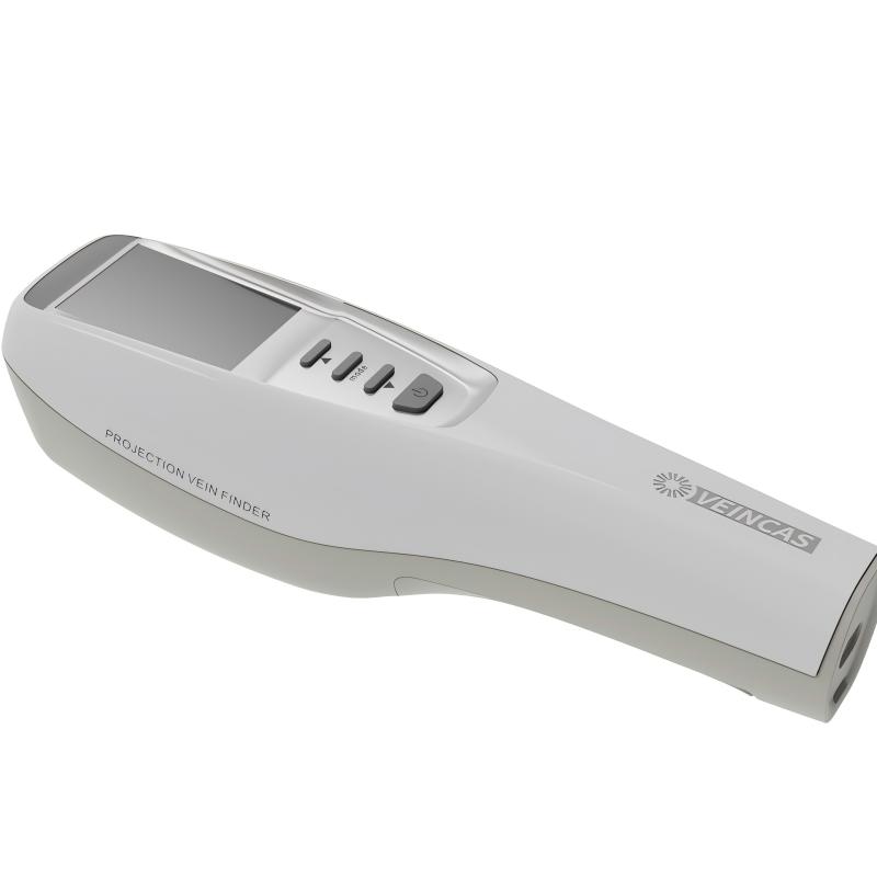
A Projection Vein Finder provides a visual reference to help healthcare professionals identify superficial veins beneath the skin. By displaying the visible course of vascular structures, this technology may support vein assessment and puncture-site selection when conventional visual inspection and palpation provide limited information.
However, the clinical value of a Projection Vein Finder should be evaluated using evidence from the specific device, patient population, and clinical setting. Improved vein visibility does not necessarily guarantee successful cannulation or shorter procedure times.
A study published in China Modern Doctor in 2015 investigated projection infrared vascular imaging during peripheral venipuncture in children. The results provide one example of how vascular visualization may support pediatric venipuncture in patients with potentially difficult peripheral venous access [1].
The study, conducted by He Ying, Xiao Liping, and Li Rujin, included 69 pediatric patients treated at Banfu Hospital in Zhongshan, Guangdong, China, between February 2014 and March 2015 [1].
Participants were divided into two groups:
Visualization-assisted group: 38 children underwent peripheral venipuncture with assistance from a projection infrared vascular imaging device.
Conventional group: 31 children underwent venipuncture using conventional methods without vascular imaging assistance.
The study included children with factors associated with potentially difficult peripheral venous access, such as obesity, edema, burn-related scarring, or phlebitis. Participants ranged in age from 6 months to 14 years [1].
The researchers compared four outcomes between the two groups:
These outcomes provide different perspectives on the procedure. First-attempt success measures whether venipuncture succeeds on the initial attempt, while overall success reflects whether the procedure succeeds within the study's defined attempts. Complications and parental satisfaction provide additional information about the experience and outcomes reported in this particular clinical setting.
The study reported higher first-attempt and overall venipuncture success rates in the visualization-assisted group than in the conventional group [1].
|
Outcome |
Visualization-assisted group (n=38) |
Conventional group (n=31) |
|
First-attempt success |
84.21% (32/38) |
54.84% (17/31) |
|
Overall success |
94.74% (36/38) |
74.19% (23/31) |
The difference in first-attempt success was statistically significant (P = 0.008). The difference in overall success was also statistically significant (P = 0.034) [1].
Under the conditions evaluated in this study, projection infrared vascular visualization was associated with higher first-attempt and overall venipuncture success among the children included in the research.
These results are encouraging, but they should be interpreted within the study's limitations. The sample consisted of 69 children from a single hospital, and the findings reflect the particular device, clinical procedures, and patient population evaluated. They do not establish that every Projection Vein Finder will achieve the same success rates in other pediatric settings.
Repeated venipuncture attempts can increase procedural burden for children and caregivers. Identifying a suitable vein before insertion may therefore be helpful when conventional assessment is difficult.
A Projection Vein Finder can provide an additional visual reference for locating superficial veins. Nevertheless, successful cannulation depends on more than vein visibility alone. Vessel accessibility, patient movement, the clinician's experience, insertion technique, and other clinical factors may also influence the outcome.
For this reason, first-attempt success rates should be interpreted alongside the study design and the characteristics of the patients and procedures involved.
The researchers also compared complications associated with venipuncture in the two groups [1].
In the visualization-assisted group, 1 of 38 children developed phlebitis, corresponding to a reported complication rate of 2.63%.
In the conventional group, 4 children developed phlebitis and 3 experienced redness at the puncture site. The reported complication rate was 22.58%.
According to the original paper, the difference between the groups was statistically significant (P < 0.05) [1].
These findings indicate that fewer complications were observed in the visualization-assisted group in this particular study. However, they do not prove that using a Projection Vein Finder eliminates complications or is without risk.
Venipuncture outcomes may be influenced by patient characteristics, vascular condition, insertion technique, operator experience, catheter management, and subsequent care. The study's complication findings should therefore be presented as results from one defined clinical setting rather than as a universal safety claim for vascular visualization devices.
The study assessed parental satisfaction using a questionnaire developed by the hospital's nursing team. The questionnaire covered areas including psychological support, health education, and perceived proficiency in venipuncture [1].
The reported satisfaction rates were:
|
Group |
Satisfied parents |
Satisfaction rate |
|
Visualization-assisted group |
37/38 |
97.37% |
|
Conventional group |
24/31 |
77.42% |
The original paper reported a statistically significant difference between the groups [1].
These findings suggest that parental satisfaction was higher in the visualization-assisted group under the conditions of the study. However, the questionnaire was developed locally for the research, and the sample size was limited.
The results should therefore be interpreted within that context rather than generalized to all pediatric patients, caregivers, or healthcare facilities.
Near-infrared vascular imaging uses differences in the optical properties of blood and surrounding tissue to help generate an image of superficial veins. Depending on the system, the processed image may be displayed on a screen or projected onto the skin to provide a visual reference during vein assessment [2].
For pediatric venipuncture, this type of visualization may offer additional information when veins are difficult to identify through conventional inspection and palpation alone.
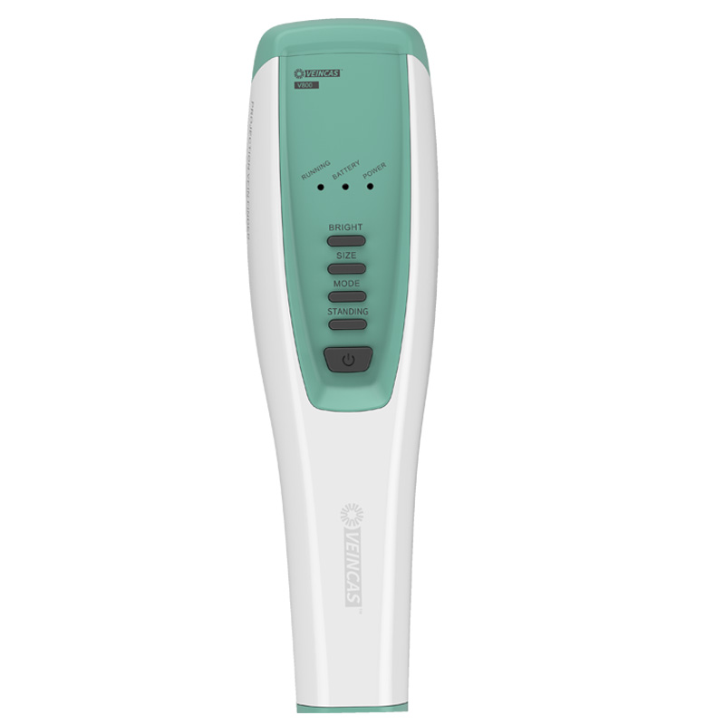
A Projection Vein Finder can provide a visual reference for assessing the visible course of superficial veins. This may help healthcare professionals identify potential puncture sites before insertion.
The image represents the information detected and processed by the device. It should not be interpreted as a complete representation of every underlying vessel or as confirmation that a visible vein is suitable for cannulation.
The vascular image may help clinicians compare potential sites alongside other clinical information, including vessel accessibility, the patient's condition, and the requirements of the planned procedure.
The final choice of puncture site should remain based on professional assessment and applicable clinical protocols.
A projected vascular image may help clinicians relate the observed vein pattern to the intended puncture site. However, the image does not replace appropriate insertion technique or established methods for assessing needle and catheter placement.
The practical value of the image depends on the specific device, its operating conditions, and how the information is incorporated into clinical practice.
The 2015 study included children with factors associated with difficult peripheral venous access and reported better puncture outcomes in the visualization-assisted group [1].
This provides a clinical example of how a Projection Vein Finder may be used during pediatric venipuncture. Further evidence is needed to determine how consistently the findings apply across different patient groups, clinical settings, and device systems.
The findings of the 2015 study should be considered alongside other clinical research. Studies of near-infrared vascular imaging have not consistently demonstrated that improved vein visibility leads to higher first-attempt cannulation success.
A cluster-randomized clinical trial published in Anaesthesia in 2013 included 1,913 children scheduled for elective surgery. The researchers evaluated three near-infrared vascular imaging devices and compared their use with conventional practice [2].
Suitable veins were more easily visible with two of the evaluated devices than with the third. However, first-attempt cannulation success did not differ significantly among the groups, with reported rates ranging from 73.1% to 75.3% (P = 0.93) [2].
This finding highlights an important distinction between image visibility and procedural success. A device may make suitable veins easier to see without necessarily increasing the probability of successful cannulation on the first attempt.
A separate cluster-randomized clinical trial published in Pediatrics in 2013 included 770 children scheduled for elective surgery [3].
First-attempt cannulation success was 70% in the group using near-infrared imaging and 71% in the conventional group. The difference was not statistically significant (P = 0.69). The study also found no statistically significant difference in the time required to obtain intravenous access [3].
The researchers noted that veins could be visualized in most patients, but the device did not improve the measured cannulation outcomes in that study.
A randomized clinical trial published in Paediatric Anaesthesia in 2014 investigated near-infrared vascular imaging in 226 children referred for difficult cannulation [4].
First-attempt success was 59% in the visualization group and 54% in the control group. The difference was not statistically significant (P = 0.41). The study also found no statistically significant difference in median time to successful cannulation [4].
This research is particularly relevant to difficult venous access because the participants had already been identified as challenging to cannulate. Nevertheless, the device did not significantly improve the measured outcomes in that study.
The studies above demonstrate that clinical outcomes can vary according to the device, patient population, and study conditions.
The 2015 study provides evidence of improved puncture outcomes in its specific pediatric population [1], while other randomized trials report that better visibility does not necessarily translate into better cannulation outcomes [2–4]. These findings should be presented together to give readers a balanced understanding of the current evidence.
Clinical research on Projection Vein Finders should be assessed according to the study design, device evaluated, patient population, and measured outcomes.
The 2015 pediatric study has several important limitations:
Study-specific outcomes: Success rates, complications, and parental satisfaction reflect the procedures and assessment methods used in the study.
Publication date: The study was published in 2015 and represents one part of the clinical evidence rather than a comprehensive assessment of all current vascular visualization technology.
Other pediatric trials have reported mixed results for first-attempt success and procedure time [2–4]. This reinforces the importance of evaluating clinical claims against evidence relevant to the specific device and intended use.
In practice, a Projection Vein Finder should be used as an adjunct to professional judgment, patient assessment, appropriate venipuncture technique, and applicable clinical protocols. Healthcare professionals should follow the device's instructions, precautions, and relevant regulatory requirements.
A 2015 study involving 69 children with potentially difficult peripheral venous access reported higher first-attempt and overall venipuncture success rates in the group using projection infrared vascular imaging than in the conventional group. The visualization-assisted group also had a lower reported complication rate and higher parental satisfaction [1].
These findings suggest a potential role for a Projection Vein Finder as an aid during pediatric venipuncture. However, other randomized pediatric studies have shown that improved vein visibility does not always lead to higher first-attempt success or shorter procedure times [2–4].
The available evidence should therefore be interpreted in the context of the device, patient population, clinical setting, and outcome measures. A Projection Vein Finder can provide additional visual information during vein assessment, but it should not be presented as a guarantee of successful insertion or as a substitute for clinical training and established venipuncture practices.
Q1: What is a Projection Vein Finder?
A: A Projection Vein Finder is a vascular visualization device that projects a processed image of superficial veins onto the skin, providing a visual reference during vein assessment.
Q2: Can a Projection Vein Finder help with pediatric venipuncture?
A: A 2015 study of 69 children with potentially difficult peripheral venous access reported higher first-attempt and overall success rates in the visualization-assisted group than in the conventional group [1]. These findings apply to the specific study and should not be treated as guaranteed results for all devices or clinical settings.
Q3: What first-attempt success rate did the 2015 study report?
A: The study reported a first-attempt success rate of 84.21% in the visualization-assisted group and 54.84% in the conventional group [1].
Q4: Does a Projection Vein Finder guarantee successful venipuncture?
A: No. It provides additional visual information but does not guarantee successful cannulation. Patient characteristics, vein accessibility, operator technique, and clinical conditions may also affect the outcome.
Q5: Does improved vein visibility always improve cannulation success?
A: No. Several randomized pediatric studies have reported improved vein visibility without a statistically significant improvement in first-attempt cannulation success [2–4].
Q6: Does a Projection Vein Finder replace conventional vein assessment?
A: No. It should be used alongside professional judgment, patient assessment, appropriate insertion technique, and established clinical protocols.
[1] He Y, Xiao LP, Li RJ. Effect of projection infrared vessel imager for peripheral venous puncture in children. China Modern Doctor. 2015;22(31):192–194. Chinese.
[2] de Graaff JC, Cuper NJ, Mungra RAAM, Vlaardingerbroek K, Numan SC, Kalkman CJ. Near-infrared light to aid peripheral intravenous cannulation in children: a cluster randomised clinical trial of three devices. Anaesthesia. 2013;68(8):835–845. doi:10.1111/anae.12294.
[3] Cuper NJ, de Graaff JC, Verdaasdonk RM, Kalkman CJ. Near-infrared imaging in intravenous cannulation in children: a cluster randomized clinical trial. Pediatrics. 2013;131(1):e191–e197. doi:10.1542/peds.2012-0968.
[4] de Graaff JC, Cuper NJ, van Dijk ATH, Timmers-Raaijmaakers BCM, van der Werff DBM, Kalkman CJ. Evaluating NIR vascular imaging to support intravenous cannulation in awake children difficult to cannulate: a randomized clinical trial. Paediatric Anaesthesia. 2014;24(11):1174–1179. doi:10.1111/pan.12501.
Leave A Message
Scan to WhatsApp :


 IPv6 network supported