 English
 Français
 Español
 Deutsch
 Italiano
 العربية



Tel :+86-755-86961139
Email :sales@vivo-light.com


Varicose veins are a common manifestation of chronic venous disease. They occur when venous valves do not function properly, allowing blood to flow backward and accumulate in superficial veins. As the condition progresses, patients may experience visible enlarged veins, leg discomfort, swelling, heaviness, and other symptoms that can affect daily activities.
Modern varicose vein management includes several treatment options, ranging from conventional surgery to minimally invasive procedures such as endovenous ablation and foam sclerotherapy. Alongside these treatment methods, vascular visualization technology can provide clinicians with an additional way to identify superficial veins before and during certain procedures.
Projection-type vein visualization systems use near-infrared imaging to detect superficial blood vessels and project their positions onto the skin. This non-contact approach may help clinicians identify visible vein pathways and plan treatment more conveniently. This article explores how projection vein visualization can support minimally invasive varicose vein treatment, drawing on clinical research published in 2017 and examining how modern vein visualization technology fits into today's clinical workflows.
Traditional surgical treatment for varicose veins commonly involves high ligation of the great saphenous vein and removal of affected superficial veins through small incisions. Although this approach can be effective for appropriately selected patients, it involves surgical incisions and may require a longer recovery period compared with some minimally invasive procedures.
Foam sclerotherapy is another treatment option. It involves injecting a prepared sclerosant foam into an affected vein. The foam contacts the inner vein wall, triggering a controlled reaction that causes the vessel to close over time. The treated vein is subsequently absorbed or incorporated into surrounding tissue through the body's healing process.
In clinical practice, treatment selection depends on the patient's symptoms, venous anatomy, reflux patterns, ultrasound findings, and overall health. Current clinical guidelines emphasize duplex ultrasound assessment when diagnosing varicose veins and planning treatment. Foam sclerotherapy may be considered for selected patients and vein types, but it is not a universal replacement for surgery or other endovenous treatments.
For procedures involving superficial veins, identifying the vessel's location before injection is an important practical step. Projection vein visualization technology offers a non-contact imaging reference that may complement the clinician's examination and established ultrasound-based assessment.
A clinical study published in Systems Medicine in May 2017 examined the use of a projection-type vascular imaging device to assist with non-contact vein localization during lauromacrogol foam sclerotherapy for lower-extremity varicose veins.
The study included 60 patients admitted between October 2015 and October 2016. They were randomly divided into two groups, with 30 patients in each group.
The control group received conventional high ligation of the great saphenous vein combined with stab phlebectomy. The observation group received high ligation under local anesthesia, with a projection-type vein imaging device used for non-contact localization, followed by lauromacrogol foam sclerotherapy.
The researchers compared several clinical outcomes between the two treatment groups. The reported results were as follows:
| Evaluation Indicator | Conventional Treatment Group | Combined Treatment Group |
|---|---|---|
| Reported effective rate | 66.67% | 96.67% |
| Reported complication incidence | 20.00% | 3.33% |
| Aesthetic evaluation | 63.33% | 93.33% |
| Average operation time | 56.15 ± 5.12 min | 23.47 ± 2.06 min |
| Average hospital stay | 10.01 ± 3.17 days | 4.25 ± 1.38 days |
| Average blood loss | 33.64 ± 5.02 mL | 9.76 ± 1.64 mL |
| Time to early ambulation | 3.45 ± 0.68 h | 0.94 ± 0.20 h |
The study reported statistically significant differences between the two groups across the evaluated indicators. The combined treatment group had a shorter average operation time and hospital stay, lower reported blood loss and complication incidence, and a higher reported effective rate and aesthetic evaluation.
However, these findings should be interpreted in the context of the study design. The researchers compared two different treatment protocols, not the same procedure performed with and without a vein visualization device. The observation group received a combination of local-anesthesia surgery, projection-based vein localization, and foam sclerotherapy. Therefore, the reported differences cannot be attributed to the imaging device alone.
Projection vein visualization systems generally use near-infrared light to help identify superficial blood vessels. Hemoglobin in the blood absorbs near-infrared light differently from surrounding tissues. A camera and image-processing system can detect these differences and generate a visual representation of the underlying superficial veins.
The processed image is then projected back onto the skin, allowing the clinician to see a surface reference corresponding to the detected vein pathway. Because the system does not require direct contact with the skin to display the vascular image, it can be used as a supplementary visualization method during certain clinical procedures.
The 2017 clinical study described a projection-type vascular imaging device operating at 850 nm and 940 nm wavelengths. It reported that the device could display superficial blood vessels located at approximately 10 mm beneath the skin. These specifications belong to the equipment described in that particular study and should not be treated as universal specifications for every vein visualization system.
In practical use, the visibility of superficial veins can vary according to vessel depth, diameter, blood flow, skin characteristics, ambient lighting, and other factors. Projection imaging therefore provides an additional visual reference rather than a complete assessment of the venous system.
Foam sclerotherapy involves introducing a sclerosant foam into a target vein. Compared with a liquid formulation, foam can displace blood within the treated vessel and increase contact between the sclerosant and the vein wall. The resulting endothelial reaction can lead to vessel closure.
In the 2017 study, the researchers used lauromacrogol, also known as polidocanol, to prepare the foam. The reported preparation involved mixing 2 mL of 1% lauromacrogol with 8 mL of air using the Tessari technique. The foam was then injected into selected superficial veins following vein localization.
Projection-based visualization was used to help identify the superficial vein pathway before injection. This can provide clinicians with a visible surface reference when locating target vessels. The actual injection process, however, still requires appropriate clinical judgment, knowledge of venous anatomy, and suitable patient assessment.
It is also important to distinguish surface vein visualization from duplex ultrasound. A projection vein finder is designed to display superficial vessels, while duplex ultrasound can evaluate deeper venous structures, blood flow, and reflux. These technologies serve different purposes and should not be considered interchangeable.
For this reason, non-contact vein visualization is best understood as a supplementary tool within a clinician-directed treatment workflow. It does not replace preoperative ultrasound assessment, appropriate treatment planning, or ultrasound guidance when clinically indicated.
The 2017 study provides an example of how projection vein imaging was incorporated into a combined treatment approach for lower-extremity varicose veins. Its findings also highlight several practical considerations when evaluating visualization technology in clinical workflows.
More visible superficial vein pathways: Projection imaging can make selected superficial veins easier to identify on the skin. This may help clinicians understand the visible distribution of target vessels before performing certain procedures.
A non-contact visualization process: Since the vascular image is projected onto the skin, the system can provide a visual reference without requiring the imaging unit to touch the patient's skin. This may be useful when clinicians need to observe superficial vein pathways during treatment preparation.
Support for procedure planning: A clearer surface reference may help clinicians coordinate vein localization with the planned treatment approach. The final treatment decision and injection technique must still be based on the patient's clinical condition and appropriate diagnostic findings.
Potential workflow advantages: The combined treatment group in the study had shorter reported operation times and hospital stays. These results are encouraging for the treatment protocol evaluated, but they do not establish that projection imaging alone produces the same benefits.
Overall, the study illustrates the potential role of projection vein visualization as one component of a minimally invasive treatment workflow. Larger studies with clearly defined device models, comparison groups, and longer follow-up periods would be useful for evaluating its independent clinical contribution.
Although the findings of the 2017 study are relevant to the discussion of projection vein visualization, several limitations should be considered before applying its conclusions more broadly.
First, the study included only 60 patients from a single hospital. A larger, multicenter study would provide a stronger basis for evaluating whether similar outcomes can be observed across different patient populations and clinical settings.
Second, the two groups received different treatment combinations. Since the observation group received local-anesthesia surgery, projection-based localization, and foam sclerotherapy, the study cannot isolate the effect of the imaging device from the effects of the other treatment components.
Third, the paper did not provide long-term follow-up data. Varicose vein treatment outcomes may change over time, and recurrence, vein closure, symptom improvement, and other clinical endpoints require appropriate follow-up assessment.
These limitations help define what the study can reasonably demonstrate: projection-based vein localization was used as part of a combined treatment protocol, and the overall protocol was associated with better short-term outcomes than the conventional treatment approach evaluated in that study.
As near-infrared imaging technology continues to develop, projection vein visualization systems are becoming increasingly useful in clinical environments where superficial vascular visibility is important. Improvements in image processing, vein mapping, depth indication, and visualization modes provide healthcare professionals with additional options for observing superficial blood vessels.
Vivolight develops vessel visualization systems designed to help healthcare professionals identify superficial veins through projected near-infrared imaging. These systems provide non-contact visual references that can support vein observation in different clinical scenarios.
For example, the V900P Projection Vein Finder offers real-time vein mapping, depth indication, and multiple visualization options. These features help users observe superficial vein pathways and distinguish veins at different estimated depths. Such functions can be useful when superficial vein visibility is an important part of clinical preparation or assessment.
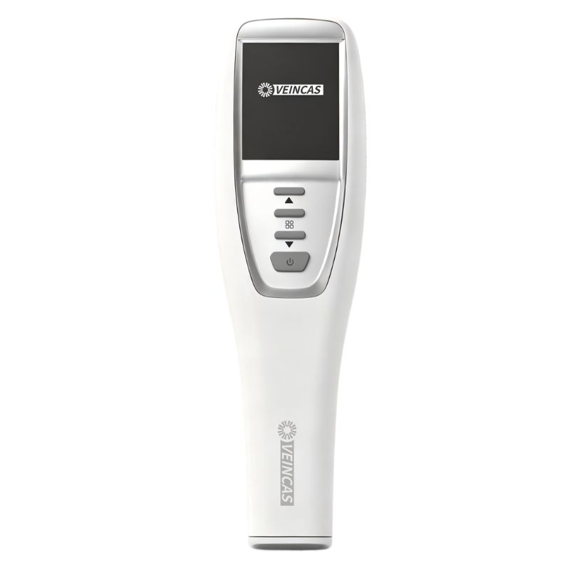
Vivolight V900P Projection Vein Finder
The V800P Vessel Visualization System uses 850 nm near-infrared imaging and offers an investigation depth of up to 10 mm according to its product catalogue. It also provides different image resolution and visualization options to accommodate various vein observation requirements.
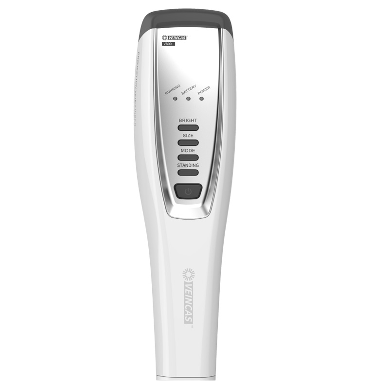
V800P Projection Vein Finder
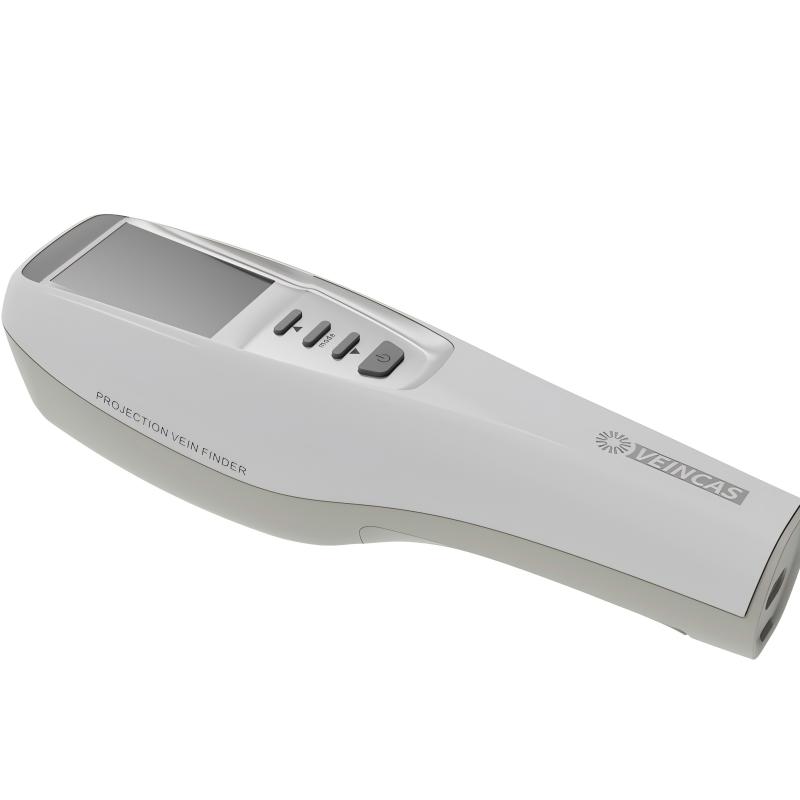
V900P Product Design and Imaging Device
Beyond varicose vein-related applications, projection vein visualization technology can also support other clinical scenarios where superficial veins need to be identified. These include venipuncture, cosmetic procedures, and selected vascular procedures.
Healthcare providers evaluating these systems should consider factors such as imaging depth, image clarity, vein detection capabilities, visualization modes, and ease of operation. The appropriate equipment depends on the intended clinical application and the requirements of the healthcare facility.
Through its Projection Vein Finder product range, Vivolight provides different equipment options for healthcare professionals seeking practical near-infrared vein imaging solutions. Understanding the capabilities of each model can help clinical teams select a system that fits their workflow while maintaining appropriate diagnostic and treatment standards.
Leave A Message
Scan to WhatsApp :


 IPv6 network supported