 English
 Français
 Español
 Deutsch
 Italiano
 العربية



Tel :+86-755-86961139
Email :sales@vivo-light.com


Placing an iv line in children often presents significant medical challenges for clinicians. Unique anatomical features like small vessel size and baby fat obscure peripheral veins. High stress levels in children cause sudden vessel constriction. Consequently, approximately 20% to 25% of pediatric patients possess specific risk factors that complicate peripheral intravenous catheter placement, classifying them as difficult iv access cases. Caregivers understandably feel anxious during these painful iv procedures. However, achieving successful pediatric intravenous access requires specialized clinical skills alongside modern visualization tools like Vivolight technology. These advanced non-invasive solutions help medical staff locate delicate vessels quickly, easily, and very accurately.
[Source: Comparative clinical data benchmarks from randomized controlled trials (RCTs) evaluating pediatric difficult intravenous access (DIVA) rates (20%–25%) and advanced vascular access modalities (Ultrasound vs. Infrared Visualization), published in Annals of Emergency Medicine.]
Children have tiny blood vessels and extra fat layers that hide delicate veins.
Stress and fear cause pediatric veins to shrink and collapse during procedures.
Movement from anxious young patients lowers first-attempt success rates for nurses.
Modern vein finder devices map hidden vessels on the skin to reduce painful needles.
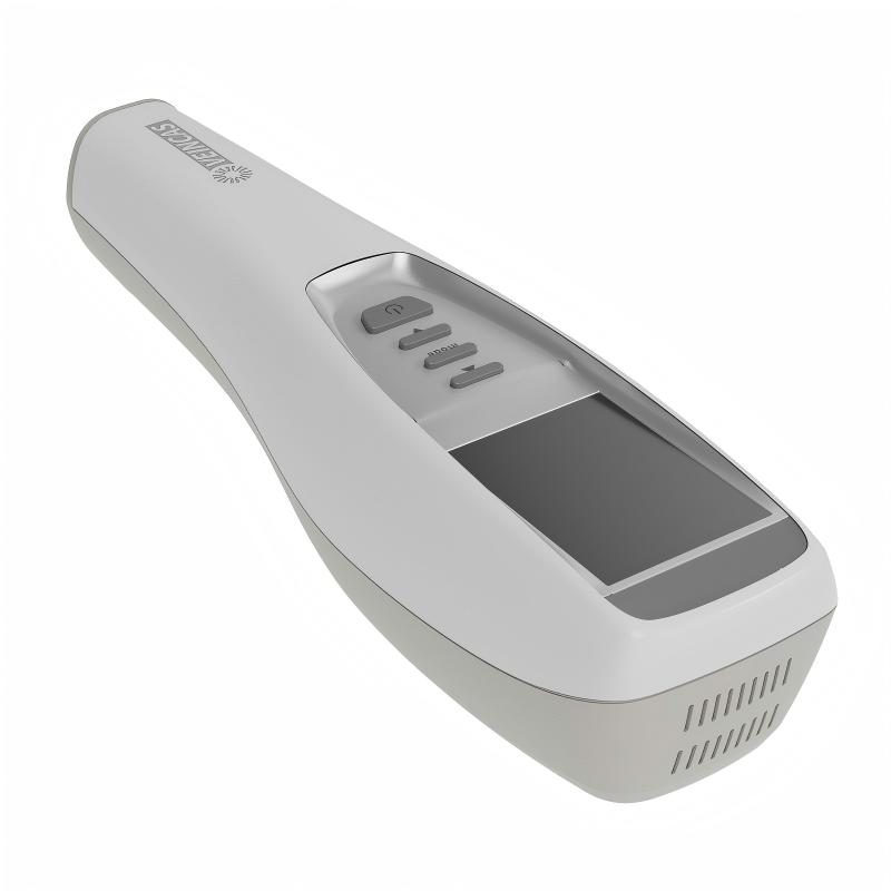
Medical providers frequently face difficult intravenous access when treating young patients in hospital settings. Clinicians define difficult intravenous access as a condition where standard peripheral catheter insertion requires elevated effort or specialized equipment. Adult patients usually have larger, well-anchored peripheral blood vessels. In contrast, children present significantly higher failure rates during initial catheter placement procedures.
Frequent failure during initial cannulation leads to multiple iv attempts, causing physical pain and psychological trauma for children. Repeated needle punctures damage peripheral veins rapidly. Healthcare teams must recognize difficult iv access early to prevent medical delays and unnecessary patient distress.
Age serves as one of the most critical risk factors for iv access in pediatric patients. Developmental characteristics in infants and toddlers directly increase procedural complexity. Clinical research demonstrates a clear mathematical relationship between patient age and catheter placement success rates.
[Source: Peer-reviewed clinical trial data on pediatric intravenous access risk factors (PMID: 26109542, PMID: 31379251), evaluating the inverse mathematical correlation between age metrics and difficult IV access risks (OR: 0.91–0.94, p < 0.05).]
|
Study Source |
Age Factor Metric |
Odds Ratio (OR) |
95% Confidence Interval (CI) |
p-value |
Clinical Finding |
|---|---|---|---|---|---|
|
PMID 26109542 |
Increasing Age |
0.94 |
0.89 – 0.99 |
0.017 |
Higher age lowers the odds of experiencing difficult iv access, whereas younger age raises the likelihood. |
|
PMID 31379251 |
Age |
0.91 |
0.85 – 0.98 |
0.01 |
Age is inversely correlated with difficult intravenous access risks. |
The data confirms that younger pediatric patients experience much higher odds of challenging iv placement. Infant anatomy contains tiny superficial veins surrounded by dense subcutaneous fat layers. Smaller blood vessels make traditional visual inspection tough for pediatric nurses. Modern healthcare systems adopt high-precision devices like the Vivolight handheld vein finder to assist clinicians. This portable vein finder projects target vascular maps directly onto skin, establishing successful intravenous access safely.
Unique structural characteristics in early human development heavily influence iv access in pediatric patients. Young patients possess distinct anatomical features that change rapidly during growth. Clinicians must adapt their approach because standard adult procedures rarely work for tiny, growing bodies. Understanding these physiological structures helps medical teams anticipate obstacles during clinical routines.
Vessel caliber represents a primary obstacle during pediatric catheter placement. Infants and young children possess peripheral blood vessels with extremely small internal diameters. An adult peripheral vein typically accommodates standard catheters easily. Pediatric vessels often measure only a fraction of a millimeter in diameter. These tiny dimensions significantly reduce the margin of error during vein identification and cannulation procedures.
Smaller vessel diameters make standard iv needles appear proportionally massive. Inserting a needle into a miniature blood vessel requires exceptional precision and hand stability. A slight misalignment can puncture the opposite vessel wall, causing immediate localized bleeding or tissue infiltration. Smaller and fragile veins also limit the selection of insertion sites, forcing healthcare providers to target delicate areas like the back of the hand or the foot.
A high ratio of subcutaneous baby fat further complicates peripheral intravenous access in infants and young children. Infant limbs naturally feature thick layers of superficial fat tissue covering peripheral blood vessels. This dense adipose layer creates a physical barrier that hides deep peripheral veins from standard visual inspection.
|
Patient Group |
Subcutaneous Fat Depth |
Peripheral Vein Visibility |
Palpation Difficulty |
|---|---|---|---|
|
Infants (0–12 months) |
High thickness |
Poor visual detection |
High resistance to touch |
|
Toddlers (1–3 years) |
Moderate thickness |
Partial visual detection |
Moderate resistance to touch |
|
Adults |
Low relative thickness |
High visual detection |
Low resistance to touch |
The fat layer distorts direct tactile feedback during manual examination. Clinicians cannot easily feel underlying venous bounce through dense tissue. Consequently, healthcare providers struggle to determine accurate vessel orientation without technological assistance. Advanced tools like a Vivolight projection vein finder help clinicians project real-time vascular maps directly onto skin surface layers, bypassing heavy subcutaneous fat obstacles seamlessly.
Pediatric blood vessels feature thin, delicate structural walls that lack mature connective tissue support. These elastic walls expand and collapse rapidly under fluid pressure changes. Furthermore, the structural fragility makes blood vessels susceptible to mechanical damage during needle advance. A standard metal needle can easily shear delicate pediatric vascular linings.
|
Vessel Characteristic |
Pediatric Vascular Impact |
Clinical Effect During IV Attempt |
|---|---|---|
|
Thin Tunica Media |
Vessel walls tear or rupture under pressure |
Increased risk of blown veins during iv insertion |
|
Low Vascular Anchor |
Veins roll away from incoming needles |
Catheter tip misses target lumen during cannula insertion |
When clinicians attempt cannula insertion, fragile pediatric veins often blow or collapse immediately. The needle tip ruptures the delicate wall before proper catheter placement occurs. Additionally, unanchored peripheral veins roll easily under skin pressure. Modern tools like a portable vein finder, handheld vein finder, or dedicated pediatric vein visualization device offer real-time assistance. Medical teams utilize a pediatric vein finder or specialized vein finder for pediatric patients to overcome deep depth obstacles and secure stable intravenous access efficiently.
Physical stress drastically alters vascular conditions in sick children during acute hospital visits. Dehydration and low blood pressure reduce vein turgor and vessel visibility significantly. These physiological states cause peripheral blood vessels to collapse rapidly.
Stress triggers systemic release of adrenaline in young patients. Adrenaline causes peripheral vasoconstriction, which narrows vessel lumens quickly. Consequently, clinicians encounter extreme difficulty while securing venous access during medical emergencies.
Psychological distress directly impairs clinical performance during catheter placement. Fear triggers immediate physical resistance and uncontrollable body movement in hospitalized children.
[Source: Observational clinical study data evaluating the impact of pediatric psychological distress and behavioral factors (distress, movement, and non-compliance) on catheterization hurdles and first-attempt IV success rates.]
|
Behavioral Factor |
Specific Manifestation |
Impact on Pediatric IV Insertion |
|---|---|---|
|
Distress |
Crying, fear, and stress |
Complicates catheterization and creates procedural hurdles for 24.8% of staff |
|
Movement |
Restlessness during insertion |
Compromises procedural precision and lowers first-attempt success rates |
|
Lack of Cooperation |
Non-compliance and resistance |
Hinders essential nurse-patient dynamics required for successful entry |
Patient movement severely compromises target precision during initial needle entry. Sudden physical shifts cause vascular trauma and force clinicians into multiple iv attempts. Implementing effective procedural pain management helps calm anxious children during difficult intravenous access procedures.
Advanced medical photoelectric and laser visualization technology helps medical staff locate difficult veins non-invasively during challenging procedures. Modern optical tools illuminate subcutaneous vascular structures instantly to support successful cannulation without causing extra patient discomfort.
[Source: Comparative clinical evaluation data on first-try IV cannulation success rates in pediatric patients with difficult intravenous access (DIVA Score > 4), comparing near-infrared vein visualization technology (VeinViewer) against traditional placement methods (58% vs. 25%).]
|
Technology Used |
Patient Group |
First-Try Success (Near-Infrared) |
First-Try Success (Traditional Method) |
|---|---|---|---|
|
VeinViewer |
Pediatric DIVA (Score > 4) |
58% |
25% |
Clinicians deploy the Vivolight projection vein finder to map target vessels accurately while securing intravenous access in emergency settings. This handheld vein finder projects real-time digital vascular patterns directly onto skin surface layers. Medical teams rely on this portable vein finder as an effective pediatric vein finder. Using a pediatric vein visualization device optimizes pediatric iv line placement efficiency. This specialized vein finder for pediatric patients streamlines complex iv insertion routines, enabling accurate line placement on the first attempt during routine iv procedures.
Pediatric intravenous access presents unique clinical challenges compared to adult care. Small vessel size, thick subcutaneous fat, and fragile vessel walls hinder direct vein identification. High stress levels trigger peripheral vasoconstriction, while fear causes sudden patient movement. These anatomical, physiological, and behavioral factors make pediatric iv placement significantly more difficult for medical staff.
Caregivers can find comfort in modern clinical advancements. Healthcare professionals undergo specialized pediatric training to manage delicate vessels gently. Furthermore, advanced diagnostic tools like the Vivolight projection vein finder help clinicians locate hidden veins quickly and accurately.
Small vessel size and thick baby fat hide delicate peripheral veins. High stress levels also trigger vessel constriction in children. These combined factors frequently lead to difficult iv access during routine hospital visits.
Medical teams utilize modern vascular illumination tools. A Vivolight projection vein finder maps subterranean veins directly onto skin surfaces. This handheld vein finder helps nurses identify optimal target sites without delay.
In pediatric care, delicate veins roll away and collapse easily. Clinicians employ a high-precision pediatric vein finder to illuminate non-visible structures instantly. Using a vein finder for pediatric patients minimizes unnecessary needle sticks effectively.
Modern optical devices streamline intravenous access through real-time vascular projection. A portable vein finder highlights target vessels before needle entry. Deploying a pediatric vein visualization device optimizes iv placement, ensuring higher success rates during an iv attempt or complex iv routine.
Leave A Message
Scan to WhatsApp :


 IPv6 network supported