 English
 Français
 Español
 Deutsch
 Italiano
 العربية



Tel :+86-755-86961139
Email :sales@vivo-light.com


Difficult peripheral venous access can present a practical challenge in clinical care, particularly in older patients and individuals whose veins are difficult to identify by conventional visual inspection and palpation.
Near-infrared (NIR) vein visualization technology has been developed to assist clinicians by making superficial veins more visible beneath the skin. Rather than replacing clinical assessment or cannulation skills, vein visualization can provide additional information about the location and course of superficial veins before and during peripheral venipuncture.
Clinical studies have investigated whether this additional visualization can improve procedural outcomes. However, the results should be interpreted in the context of the specific patient population, clinical setting, operator experience, and—importantly—the specific device evaluated.
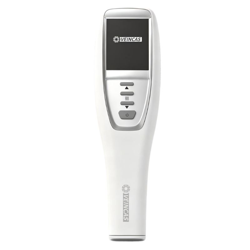
A 2019 study published in the Chinese Journal of Clinical Nursing evaluated the use of visualization-assisted venipuncture in patients treated in a cardiology department.
The study included 150 patients, with 75 patients in the conventional venipuncture group and 75 patients in the visualization-assisted group. The conventional group underwent peripheral venous cannulation using visual inspection and palpation, while the intervention group received assistance from a projection-based infrared vein imaging device.
Importantly, the study identified the device used in the intervention: projection infrared vein imaging device, manufactured by vivolight.
Both groups used 24G closed intravenous catheters manufactured by Becton, Dickinson and Company (BD), and all venipuncture procedures were performed by the same group of nurses.
The investigators reported several statistically significant differences between the two groups.
In the visualization-assisted group, 69 of 75 patients (92.0%) achieved successful venipuncture on the first attempt.
In comparison, 38 of 75 patients (50.67%) in the conventional group achieved first-attempt success.
The difference in overall venipuncture success distribution between the two groups was statistically significant (Z = −5.697, P < 0.001).
The visualization-assisted group also had fewer patients requiring multiple attempts:
These findings suggest that, in the cardiology patients evaluated in this study, visualization-assisted venipuncture was associated with a higher likelihood of successful first-attempt cannulation.
The mean venipuncture time was:
The difference was statistically significant (t = 13.096, P < 0.001).
This result indicates that the visualization-assisted approach was associated with shorter venipuncture time under the conditions of this study. Actual procedure time can vary according to vascular condition, operator experience, patient characteristics, and clinical workflow.
The study monitored several events following venipuncture, including extravasation, catheter occlusion, perivascular bruising, venous thrombosis, and phlebitis.
The reported overall rate of venipuncture-related adverse events was:
The difference was statistically significant (χ² = 8.027, P = 0.005).
These findings are encouraging, but they should not be interpreted as evidence that visualization technology eliminates complications. Venipuncture-related complications can have multiple causes, and the study was conducted in a relatively small, single-center population.
Patient satisfaction was also higher in the visualization-assisted group.
The proportion of patients classified as “satisfied” was:
The difference in satisfaction ratings was statistically significant (Z = −4.141, P < 0.001). The researchers used a department-developed satisfaction questionnaire with a reported Cronbach’s α of 0.887 and a content validity index of 0.920.
Because patient satisfaction is influenced by multiple factors beyond the visualization technology itself, these findings are best interpreted as an association observed in this particular clinical setting.
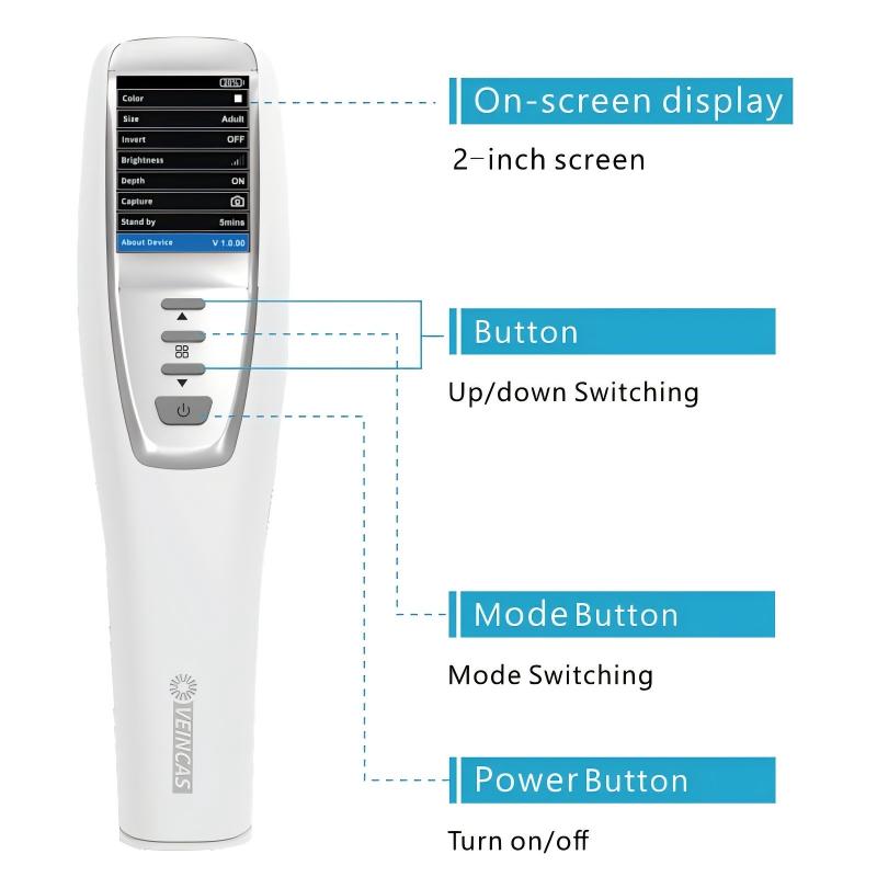
Projection-based NIR vein visualization relies on the different absorption characteristics of near-infrared light between blood hemoglobin and surrounding tissue.
The imaging system detects differences in reflected infrared light and processes the signal to produce a visual representation of superficial veins. The resulting image can help clinicians assess vein location, distribution, and course before selecting a puncture site.
In the 2019 study described above, the nurses adjusted the imaging system so that superficial veins could be clearly visualized. They assessed the visible vein pattern and selected a suitable vessel before performing peripheral IV cannulation.
This type of visualization is particularly relevant when superficial veins are difficult to identify using conventional inspection and palpation alone.
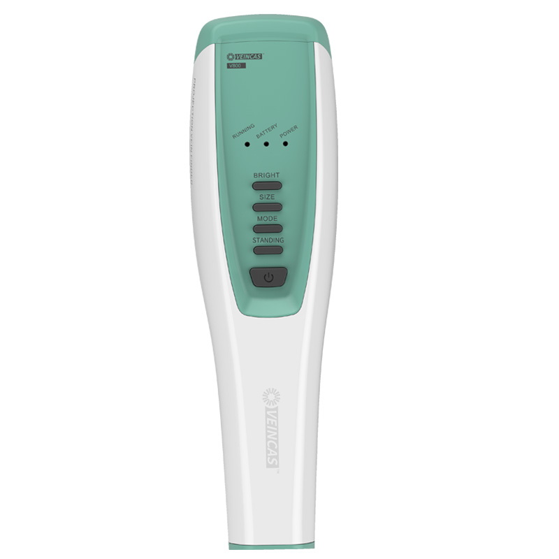
The 2019 cardiology study is not the only clinical investigation of NIR-assisted venipuncture.
For example, Fumagalli et al. conducted a pilot randomized study in elderly critically ill patients. The study enrolled 103 patients, with 47 receiving NIR-assisted venipuncture and 56 receiving standard venipuncture. The specific NIR device evaluated was EasyVein (InSono, Calenzano, Florence, Italy).
Unlike the Chinese cardiology study, this randomized pilot study did not find significant differences in procedure length, number of attempts, or reported pain. However, the NIR group had a lower incidence of hematoma: 8.5% versus 28.6% in the standard-care group (P = 0.012). The authors described the results as preliminary and indicated that further studies were needed to confirm the findings and identify which patients might benefit most.
This difference between studies is important. It illustrates why clinical evidence for vein visualization should not be reduced to a single universal claim. Outcomes can differ according to the device, study design, patient population, operator training, and clinical environment.
Taken together, the available evidence suggests that NIR vein visualization can be a useful adjunctive technology for peripheral venous access, particularly when superficial veins are difficult to locate using conventional methods.
The strongest and most appropriate interpretation is not that vein visualization guarantees successful cannulation for every patient. Instead, the technology may provide clinicians with additional visual information that can support vein assessment and puncture-site selection.
Clinical judgment remains important. Patient characteristics, vein condition, treatment requirements, catheter selection, operator experience, infection-control procedures, and institutional protocols should all be considered when performing peripheral IV access.
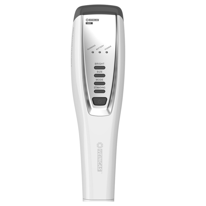
When evaluating clinical claims about vein visualization systems, it is important to distinguish between evidence for a technology category and evidence for a specific medical device.
The cardiology study discussed in this article specifically evaluated the projection infrared vein imaging device manufactured by vivolight. It should therefore be cited as evidence for that specific device and study population rather than presented as a clinical trial of every projection-based or near-infrared vein visualization system.
Similarly, other published studies have evaluated different devices, including EasyVein. Results from one device should not automatically be attributed to another device without appropriate supporting evidence.
This distinction is especially important when clinical research is used in medical-device communications.
Near-infrared vein visualization technology offers clinicians an additional method for assessing superficial veins during difficult peripheral venous access.
In a 2019 study involving 150 cardiology patients with difficult venipuncture, researchers reported differences between the visualization-assisted and conventional groups in first-attempt success, procedure time, venipuncture-related events, and patient satisfaction. These findings relate to the specific device, patient population, and clinical setting evaluated in the study.
Other clinical research has produced more mixed findings, reinforcing the importance of considering the specific device, patient population, study design, and clinical setting when interpreting the evidence.
For healthcare professionals, vein visualization technology may therefore be best understood as a visualization aid that complements—not replaces—clinical assessment and venipuncture expertise.
1. Yin D, Wang W. Application of visual operation technology in venipuncture with difficulties. Chinese Journal of Clinical Nursing. 2019;11(6):492–495. doi: 10.3969/j.issn.1674-3768.2019.06.009.
2. Fumagalli S, Torricelli G, Massi M, et al. Effects of a new device to guide venous puncture in elderly critically ill patients: results of a pilot randomized study. Aging Clinical and Experimental Research. 2017;29(2):335–339. doi: 10.1007/s40520-016-0547-0. PMID: 26914485.
3. Ke JL, Yang Y, Cao F, et al. Application effect of near-infrared visualization puncture technology in elderly patients with difficult venipuncture. Chinese Journal of Modern Nursing. 2017;23(32):4159–4161.
4. Hu JM, Yan JW. Clinical application value of vascular visualization technology in peripheral venipuncture. China Medical Equipment. 2017;14(10):137–140.
5. He Y, Xiao LP, Li RJ. Application effect of projection infrared vein imaging device in peripheral venipuncture in children. China Modern Medicine. 2015;22(31):192–194.
Leave A Message
Scan to WhatsApp :


 IPv6 network supported