 English
 Français
 Español
 Deutsch
 Italiano
 العربية



Tel :+86-755-86961139
Email :sales@vivo-light.com

A Balanced Overview of Near-Infrared Vascular Imaging: Principles, Clinical Considerations, and Technological Evolution
1. Clinical Background & Overview
The Clinical Context: Establishing peripheral intravenous (IV) access is one of the most common invasive procedures in healthcare, performed routinely across inpatient units, emergency rooms, pediatric wards, and surgical suites. In standard clinical practice, clinicians locate veins primarily through visual inspection and manual palpation. For many patients, these traditional techniques are sufficient. However, establishing venous access can become time-consuming and technically challenging in patients with Difficult Venous Access (DVA)—including young pediatric patients with small, non-prominent vessels, geriatric patients with fragile or rolling veins, individuals with higher body mass index (BMI) where veins reside deeper beneath subcutaneous fat, and patients with chronic conditions requiring frequent cannulations.
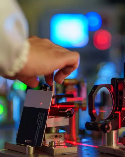
Near-infrared (NIR) vein visualization technology—often delivered via projection vein finder (PVF) devices—has been developed as an optical assistive tool to aid healthcare professionals during vascular access procedures. By utilizing the optical absorption contrast of hemoglobin in the near-infrared spectrum, these devices capture subcutaneous vascular patterns and project a real-time visual map directly onto the skin surface. This real-time visual guide is designed to assist clinicians in identifying superficial vein pathways and bifurcations prior to puncture, supporting procedural efficiency and clinical decision-making.
2. Key Takeaways
· Optical Principle: Near-infrared (NIR) vein finders utilize the differential light absorption of hemoglobin to project real-time maps of superficial veins directly onto the skin.
· Primary Clinical Value: The technology is designed to assist clinicians in locating viable superficial puncture sites, which is particularly beneficial in challenging access populations (e.g., pediatric, bariatric, and geriatric cohorts).
· Assistive Role: A vein finder provides two-dimensional superficial vascular mapping; it does not replace manual palpation, tourniquet use, vessel health assessment, or professional clinical judgment.
· Contextual Evidence: Specific published clinical trials demonstrate improvements in procedural time and first-attempt success for evaluated models (e.g., AccuVein®, VeinViewer®); however, results depend on study protocols and clinical conditions, and should not be generalized as uniform guarantees across all devices.
· Operational Safety Profile: Medical-grade projection vein finders utilize low-intensity, non-ionizing light sources engineered to comply with applicable photobiological and laser safety standards.
3. Technological Principles and System Architecture
Near-infrared vein visualization relies on the principles of optical spectroscopy and light-tissue interaction. Human skin, adipose tissue, and muscle scatter near-infrared light within the 700 nm to 1000 nm optical window, while intravascular hemoglobin (both deoxygenated and oxygenated forms) exhibits absorption peaks within this spectrum (notably around 758 nm and 929 nm).
The Step-by-Step Imaging Process
· 1. Light Emission: The device emits near-infrared light (commonly in the 750–850 nm or 850–940 nm range) onto the targeted anatomical site, penetrating superficial tissue layers up to several millimeters.
· 2. Hemoglobin Absorption: Hemoglobin flowing in subcutaneous veins absorbs the NIR light more strongly than surrounding dermal tissue, producing a natural optical contrast pattern.
· 3. Signal Capture & Processing: An integrated optical sensor equipped with specialized filtering captures the reflected NIR signals, and internal digital image processing algorithms filter noise and enhance vascular contrast.
· 4. Real-Time Skin Projection: A projection engine projects the processed vascular outline in visible light (such as green or customized contrast modes) back onto the exact anatomical location on the skin surface with minimal spatial distortion.
Comparison of Bedside Vascular Access Modalities
To select the appropriate tool, clinicians consider the anatomical requirements and operational scope of each bedside method:
|
Modality |
Primary Clinical Scope |
Key Clinical Advantages |
|
Projection Vein Finder (NIR) |
Superficial peripheral vein assessment and routine peripheral vascular access |
Projects superficial vein information onto the skin surface in real time, helping visualize vessel pathways and bifurcations; non-contact imaging |
|
Doppler Ultrasound Guidance |
Deep vessel assessment, PICC and midline catheter placement, and other vascular access procedures requiring depth information |
Provides cross-sectional vessel imaging and information on vessel depth and diameter, while supporting real-time visualization of the relative position of the needle and vessel |
|
Cold-Light Transillumination |
Superficial vessel visualization in neonates and infants |
Uses localized transillumination to assist in visualizing the location of superficial vessels |
|
Traditional Visual & Palpation |
Routine peripheral vein assessment and venipuncture |
Uses visual inspection and palpation to assess vessel location and condition without additional imaging equipment |
4. Supporting Clinical Decision-Making, Not Replacing It
In clinical vascular access, vein visualization devices are classified strictly as assistive decision-support tools. They provide visual supplementary information to enhance procedural planning, but they do not replace standard clinical assessment, tactile evaluation, or proper venipuncture technique.
Guidance from Professional Standards
The Infusion Nurses Society (INS) Standards of Practice provide guidance on assistive visualization technologies, recommending that clinicians consider the use of near-infrared (nIR) technology to facilitate the identification of viable superficial peripheral venous sites and help reduce procedural time, particularly in patients with difficult venous access or after failed initial attempts.
Anatomical Capabilities and Inherent Device Boundaries
· What the Device Shows: Displays vein trajectory, branching patterns, and bifurcations across a broad superficial field, assisting clinicians in identifying viable cannulation segments and avoiding venous valves.
· Depth & Lumen Limitations: Standard two-dimensional projection vein finders do not provide quantitative measurements of vessel depth or lumen diameter.
· Vessel Elasticity & Health: The projected image does not indicate whether a vein is sclerotic, thrombosed, fragile, or prone to rolling or collapsing upon puncture. Tactile palpation remains essential to evaluate vessel elasticity and health.
· Tourniquet Necessity: Vein visualization does not eliminate the physiological necessity of a venous tourniquet to engorge and stabilize target vessels prior to needle insertion.
5. Patient Populations That May Benefit from Visualization Support
While vein visualization can be utilized across various clinical areas, its supportive value is most apparent in patient cohorts presenting with anatomical or physiological access challenges:
· Pediatric Patients: Young children frequently possess small vessels obscured by baby fat, often accompanied by procedural movement. Near-infrared mapping provides non-invasive anatomical guidance without tactile distortion.
· Geriatric Patients: Aging can reduce skin thickness, tissue turgor, and vessel stability, making veins more prone to rolling or fragile collapse. Visual mapping assists clinicians in identifying stable vein segments and bifurcations.
· Patients with Elevated BMI: Adipose tissue scatters visible light. Near-infrared wavelengths penetrate subcutaneous fat layers to highlight superficial veins residing within standard cannulation depth.
· Chronic Disease & Oncology Patients: Patients undergoing repeated blood sampling, chemotherapy, or hemodialysis often experience progressive venous scarring. Visualization support aids in identifying viable superficial sites and supporting vascular preservation.
6. Technological Advancements in Projection Vein Imaging: The Vivolight Solution
As near-infrared vein visualization technology continues to advance, medical device manufacturers are developing more compact optical projection engines, enhanced image-processing algorithms, and adaptable display modes to meet diverse clinical environments.
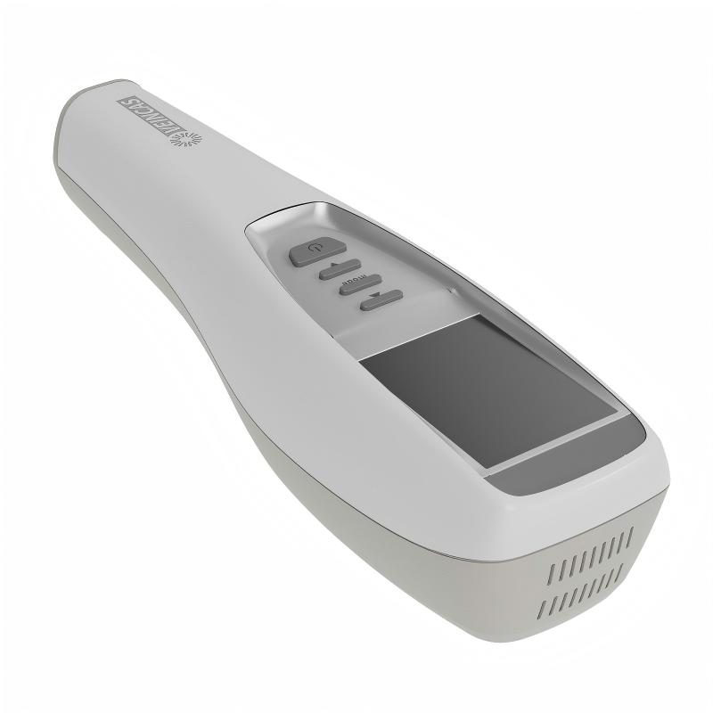
Vivolight specializes in laser-based medical imaging and projection technologies, developing dedicated visualization solutions for vascular procedures. The Vivolight V900P Projection Vein Finder utilizes advanced near-infrared imaging engineering designed to project real-time vascular outlines directly onto the patient's skin surface. Engineered to assist healthcare professionals in settings such as pediatric wards, emergency rooms, oncology departments, and general clinics, the V900P provides user-adjustable projection modes (including color customization, contrast inversion, and adjustable brightness) and optimized optical filtering. These features are designed to accommodate varying clinical lighting conditions and diverse patient skin pigmentation profiles. By providing non-contact anatomical visualization assistance before needle insertion, the V900P serves as a practical bedside tool to support clinical decision-making and peripheral vein assessment.
7. Frequently Asked Questions (FAQ)
Q1: What is the safety profile of near-infrared projection vein finders?
Medical-grade near-infrared projection vein finders utilize low-intensity, non-ionizing optical wavelengths (typically within 750–940 nm). They operate without direct skin contact, emit no ionizing radiation, and produce no thermal tissue damage when used according to manufacturer instructions. Devices are engineered to comply with applicable international laser and photobiological safety standards (such as IEC 60825-1 Class 1 and IEC 62471). As a standard operational precaution, clinicians and patients should avoid staring directly into the optical projection aperture.
Q2: Does near-infrared vein visualization work on darker skin tones?
Yes. Near-infrared imaging functions across various skin tones because the optical absorption curve of hemoglobin remains distinct from epidermal melanin within the 750–940 nm NIR window. While epidermal melanin absorbs visible light strongly, near-infrared light penetrates melanin layers to detect the contrast between blood vessels and adjacent tissue. Advanced devices also offer inverse contrast modes and customizable projection hues to further assist visibility across different pigmentation profiles.
Q3: Can a projection vein finder replace ultrasound guidance?
No. Projection vein finders and ultrasound devices serve complementary clinical roles. Projection vein finders provide wide-field, instantaneous superficial vascular mapping (<10 mm depth) for routine peripheral IV placement and venipuncture. Bedside ultrasound is necessary for deep vascular access (>10 mm), midline catheters, PICC line placement, central venous lines, and quantitative cross-sectional measurements of vessel depth and lumen diameter.
Q4: Does using a vein finder eliminate the need for tourniquets or manual palpation?
No. A projection vein finder visualizes superficial vein trajectory and bifurcations, but it cannot assess vessel elasticity, lumen patency, or internal sclerosis. Clinicians must continue applying a tourniquet to adequately engorge the vein and manually palpate the vessel to evaluate its condition before attempting cannulation.
8. References & Cited Literature
Infusion Nurses Society. (2021). Infusion Therapy Standards of Practice, 8th Edition. Journal of Infusion Nursing, 44(1S), S1-S224.
Demir, D., & Inal, S. (2019). Does the use of a vein visualization device for peripheral venous catheter placement increase success rate in pediatric patients? A randomized controlled trial. Journal of Pediatric Nursing, 44, e90-e96.
Inal, S., & Demir, D. (2017). The effect of using a vein finder on pain and first-attempt success during peripheral intravenous cannulation in children: A randomized controlled trial. Journal of Emergency Nursing, 43(4), 312-317.
Chapman, L. L., Sullivan, B., Pacheco, A. L., Draleau, C. P., & Becker, B. M. (2011). VeinViewer-assisted intravenous catheter placement in a pediatric emergency department: A randomized controlled trial. Annals of Emergency Medicine, 58(4), S262-S263.
Chiao, F. B., Resta-Flarer, F., Lesser, J., Ng, J., Ganz, A., Pino-Luey, D., ... & Wetherton, A. (2013). Vein visualization: patient characteristic factors and efficacy of a new infrared vein finder technology. British Journal of Anaesthesia, 110(6), 966-971.
Avelar, A. F. M., Peterlini, M. A. S., & Pedreira, M. L. G. (2010). Vascular visualization technologies for peripheral intravenous access in children: a systematic review. Revista Latino-Americana de Enfermagem, 18(6), 1193-1201.
Hess, H. A. (2010). A biomedical device to improve pediatric vascular access success. Pediatric Nursing, 36(5), 259-263.
Leave A Message
Scan to WhatsApp :


 IPv6 network supported